
案例 03:华东师范大学-黄正齐老师样品检测报告(鬼笔环肽·肌动蛋白)
📋 项目概况
客户单位:华东师范大学
项目负责人:黄正齐老师
项目目标:鬼笔环肽(Phalloidin)肌动蛋白(Actin)荧光标记样品的多平台检测评估,覆盖 Acti-Stain 555 Phalloidin 与 Alexa Fluor™ 532 Phalloidin 两款染料,跨 2023 年 4 月至 8 月共 6 轮检测迭代。
Reagents标记原料记录
🎨 Acti-Stain 555 Phalloidin
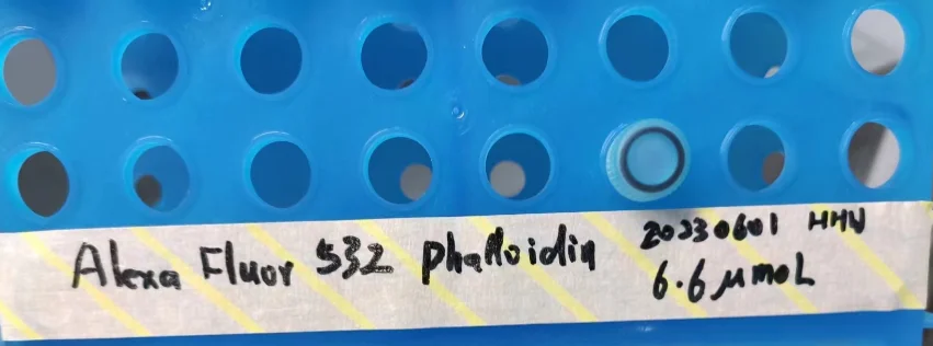
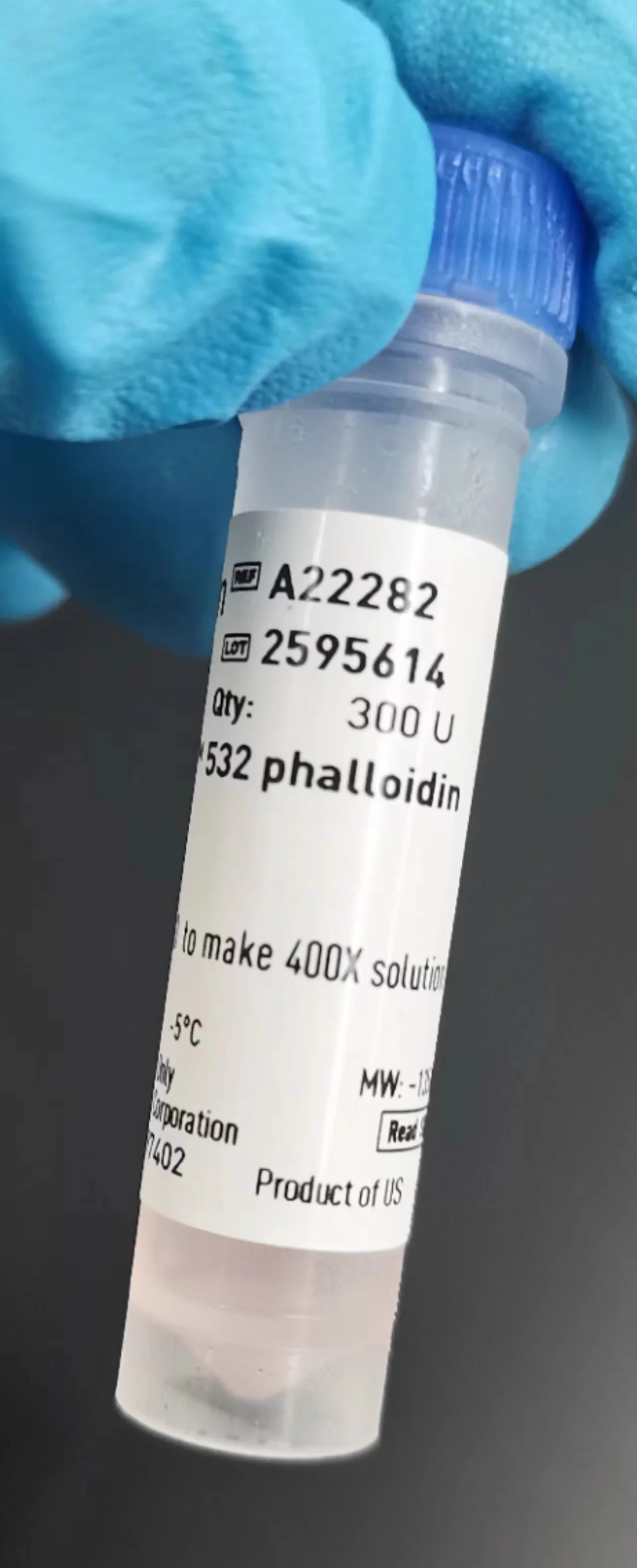
🎨 Alexa Fluor™ 532 鬼笔环肽
激发/发射波长:531 / 554 nm
产品链接:thermofisher.cn/order/catalog/product/A22282
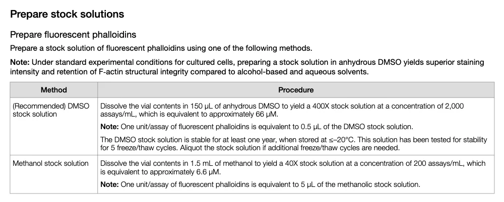
In DMSO:150 μL 66 μM stock
In methanol:1.5 mL 6.6 μM stock
本项目选择 甲醇溶解,后续需做浓度滴定(titration)以平衡信号强度与背景荧光。
使用说明 PDF:MAN0001777_Phalloidins_UG.pdf ↗
Equipment检测设备:Olympus FV3000
60× 物镜 LSM 激光光源:
20230418Actin-Alexa555 样品检测详情
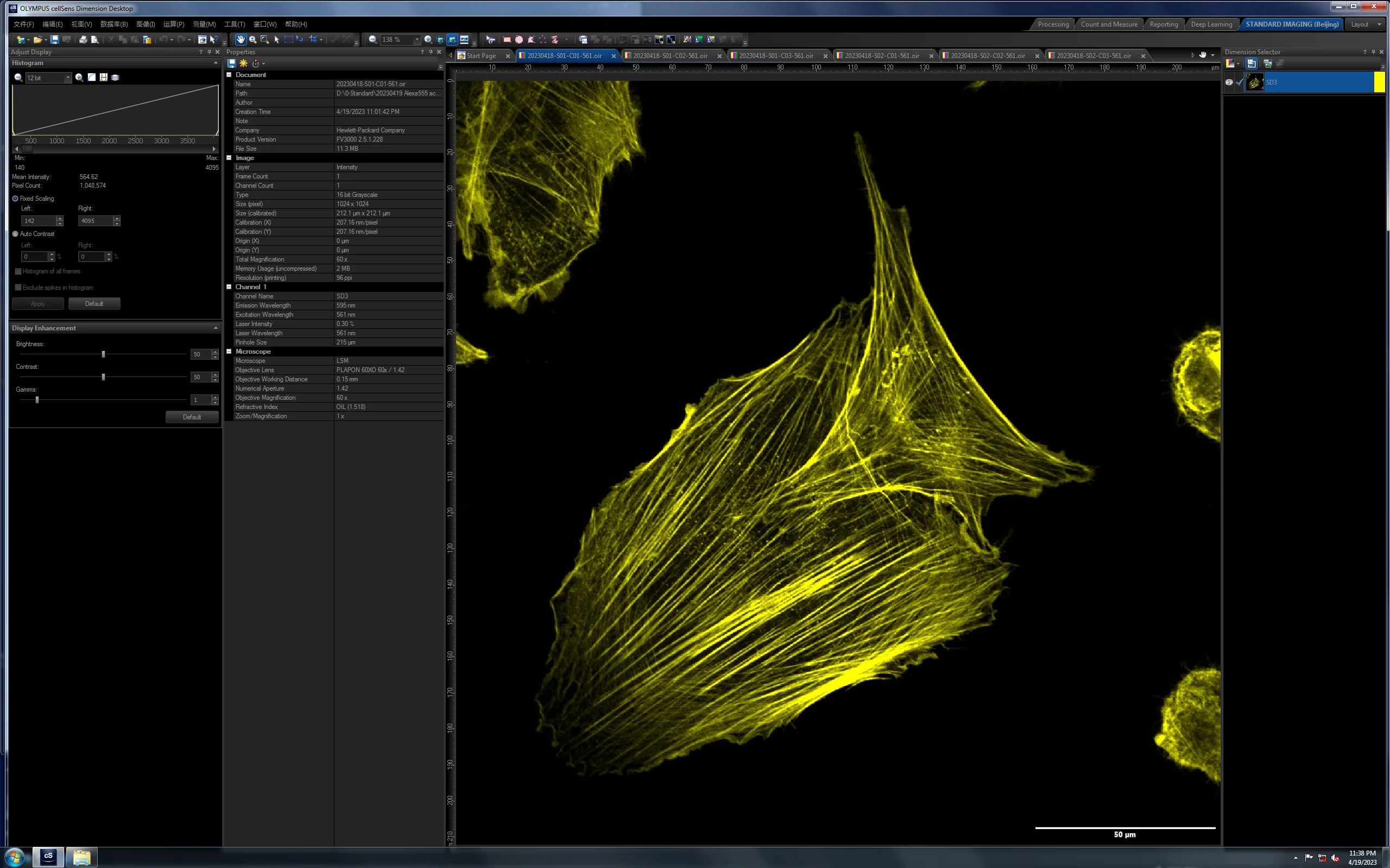
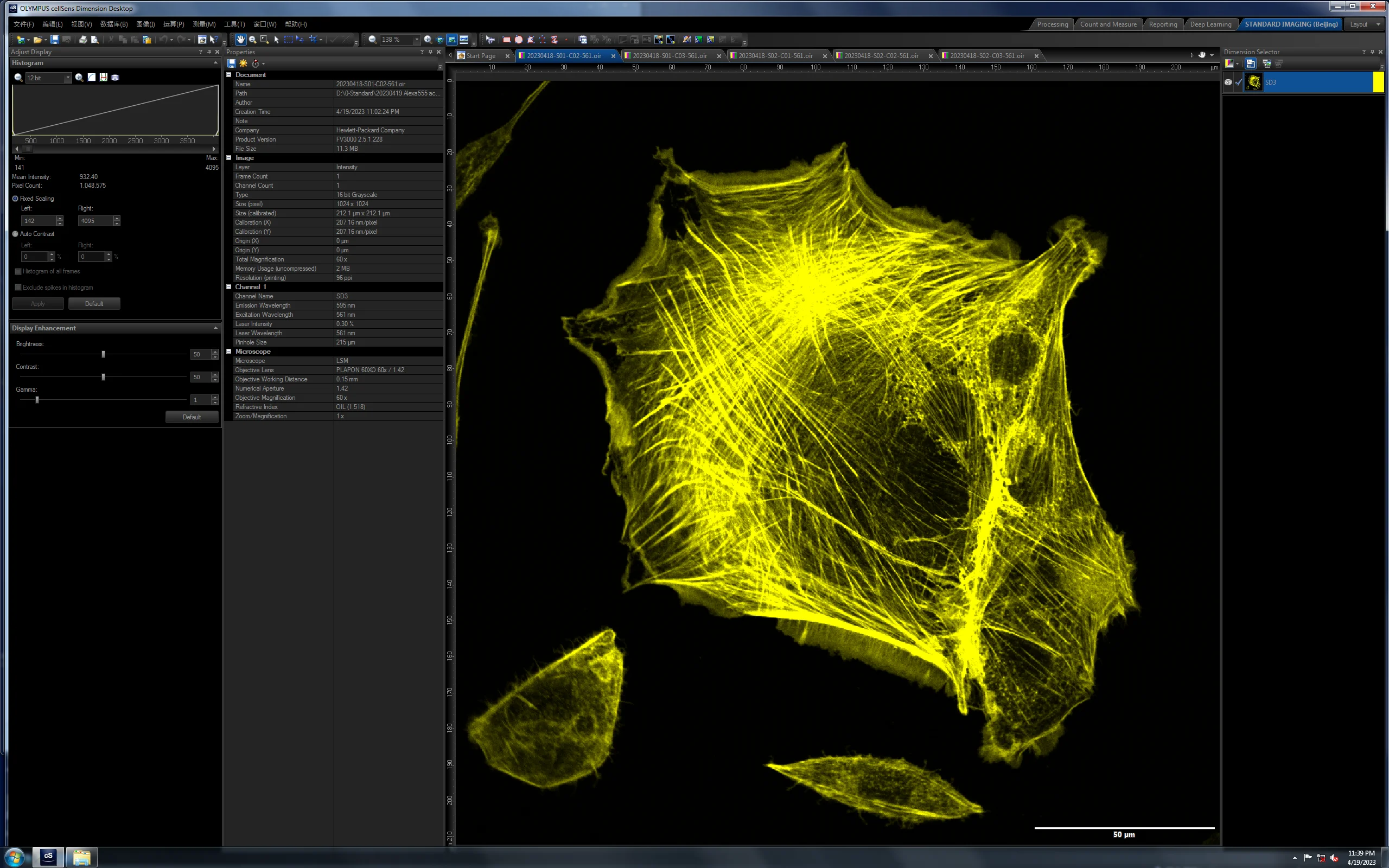
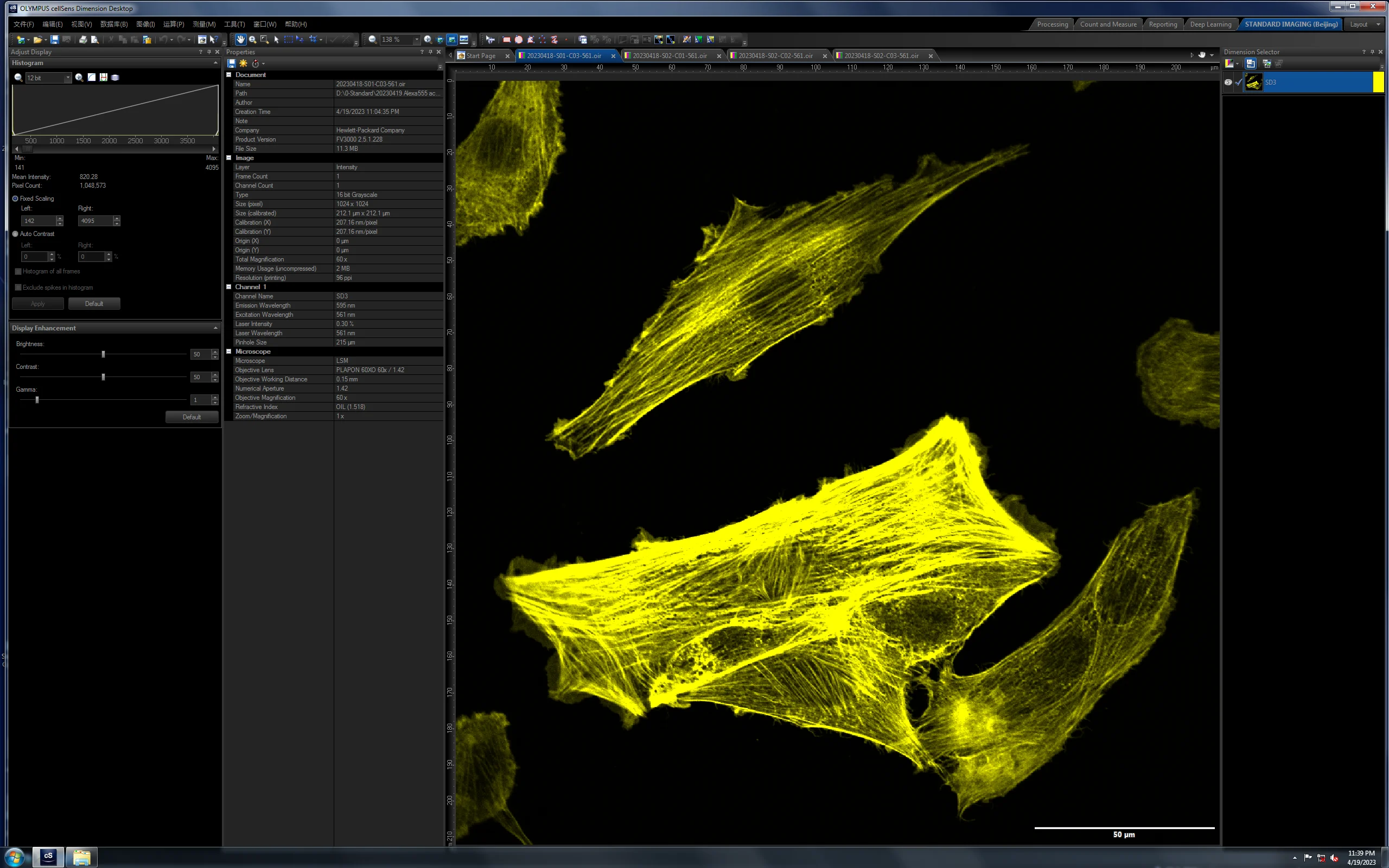
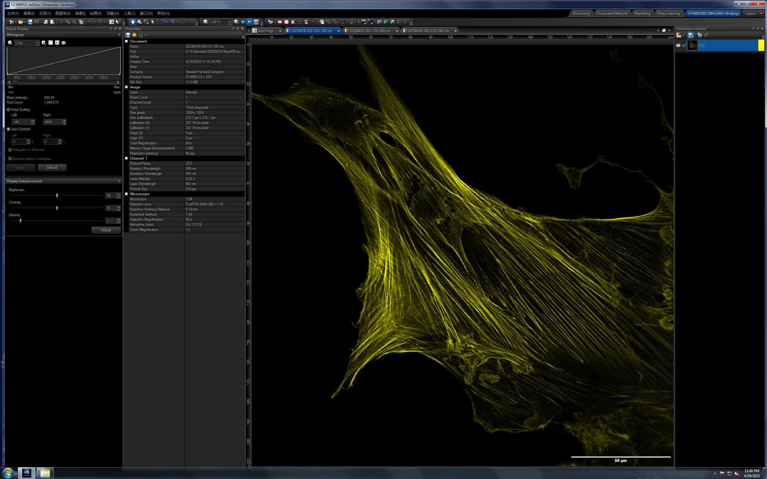
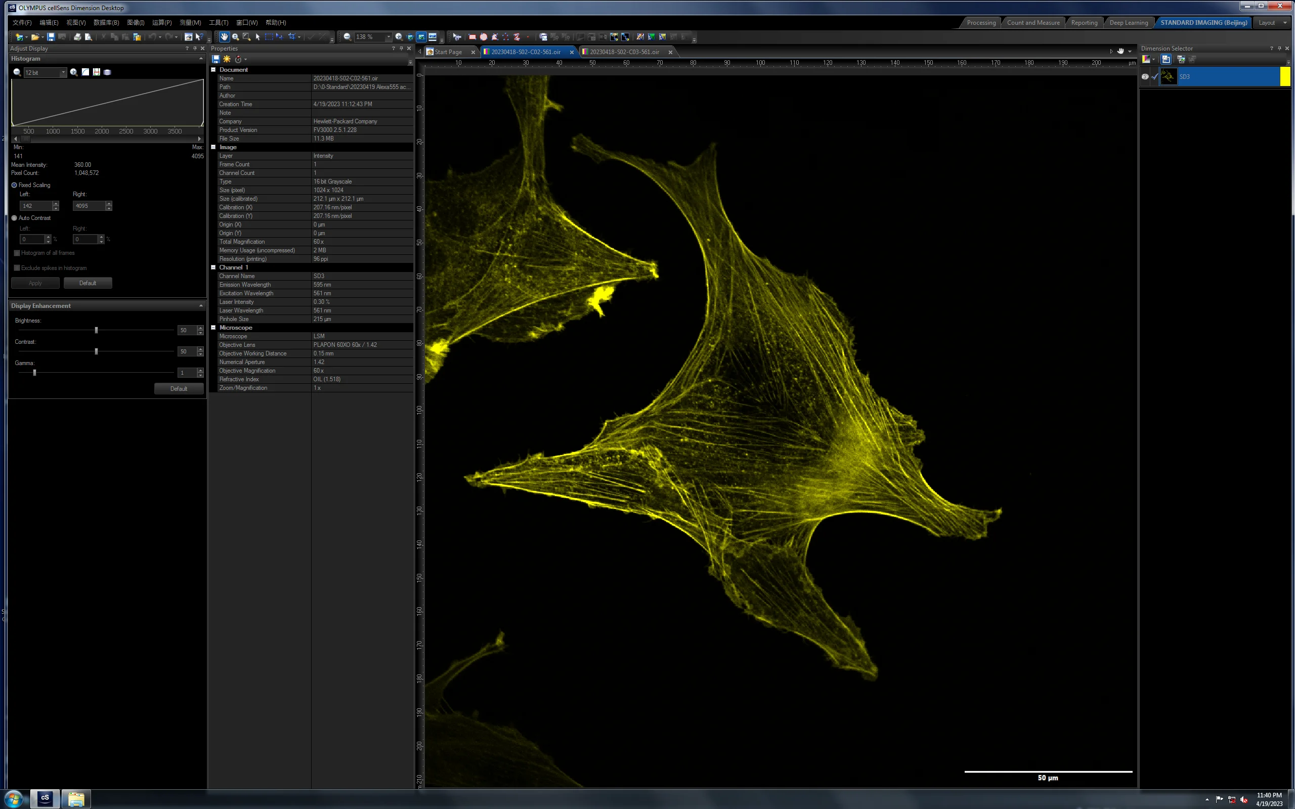
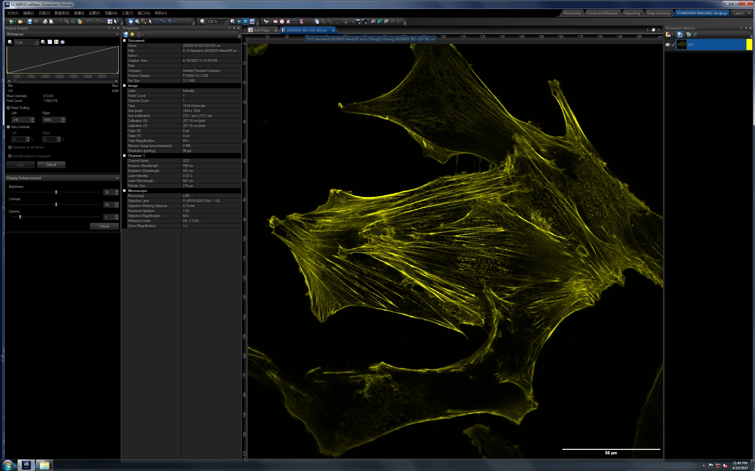
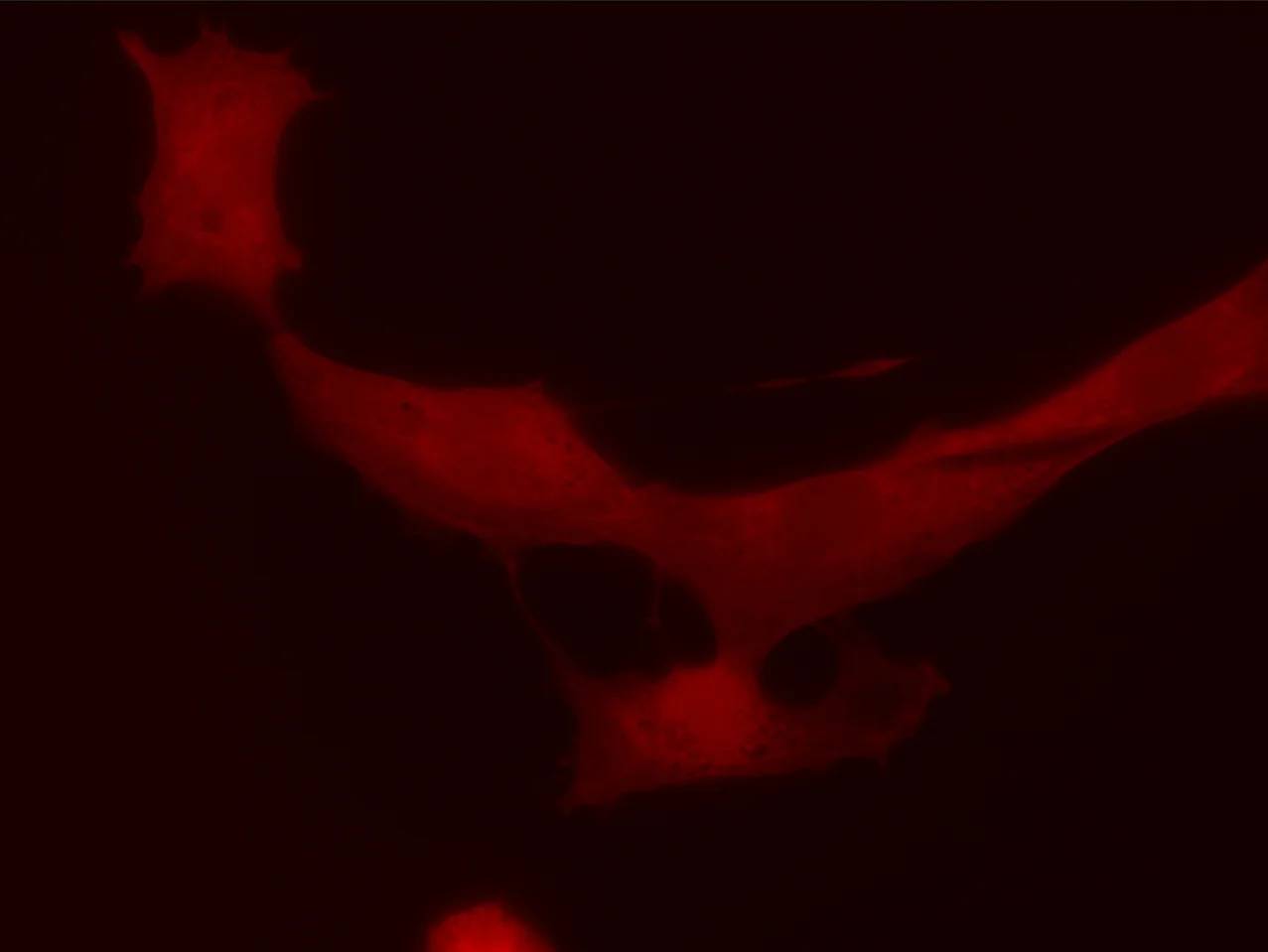
对比样品:两批 BSC-1 细胞,分别使用 4% PFA 与 4% PFA + 1% GA 两种固定方案;激光仅开 0.3%,评估低激光下两种固定剂的细节保留能力。
| 维度 | 样品 1(S01) | 样品 2(S02) |
|---|---|---|
| 标签 | S01-Alexa555 phalloidin 4% PFA Fix Bright、lowBG — 样品 A |
S02-Alexa555 phalloidin 4% PFA + 1% GA Fix Bright、higher BG、good details — 样品 B |
| 固定剂 | 4% PFA | 4% PFA + 1% GA |
| 标记信息 | Alexa555 phalloidin | Alexa555 phalloidin |
| 样品备注 | 背景更干净,雾状与丝状细节足够,样品非常明亮(0.3% 激光) | 背景较高,但信号更亮,雾状与丝状细节保存更多(0.3% 激光) |
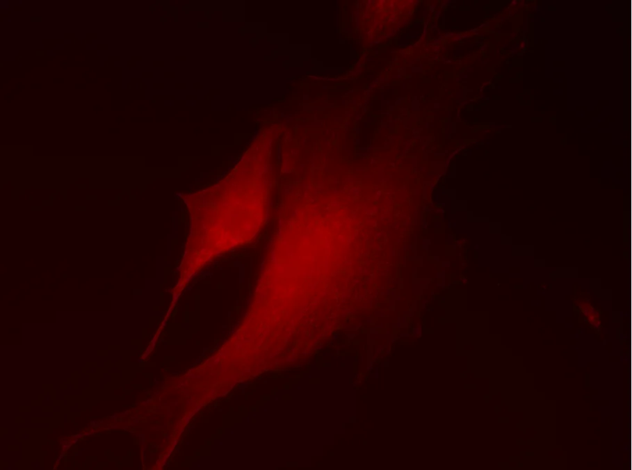
60× 共聚焦成像(S01 vs S02)
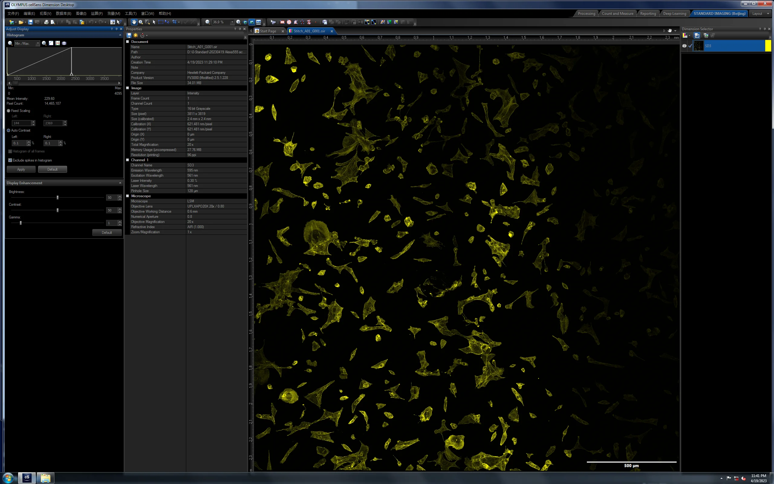
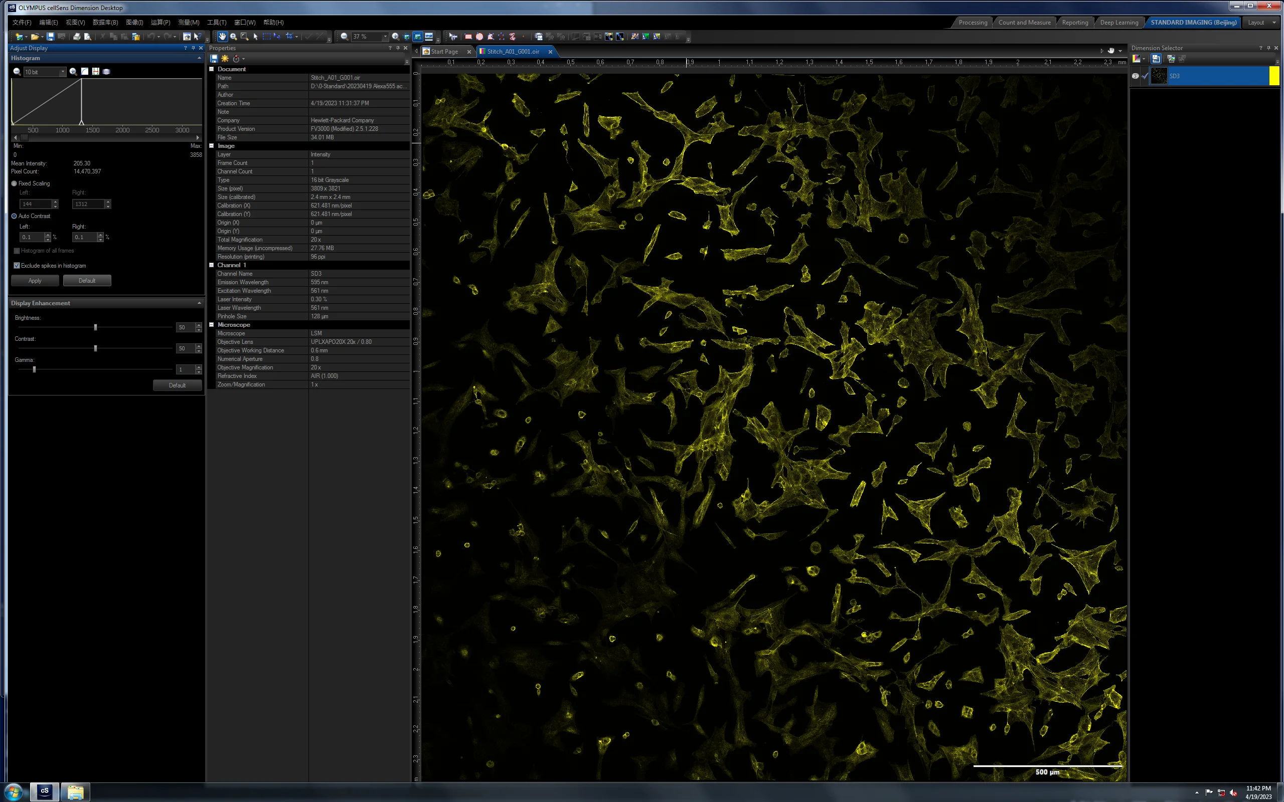
20× 低倍概览
Time扫描时间尺度
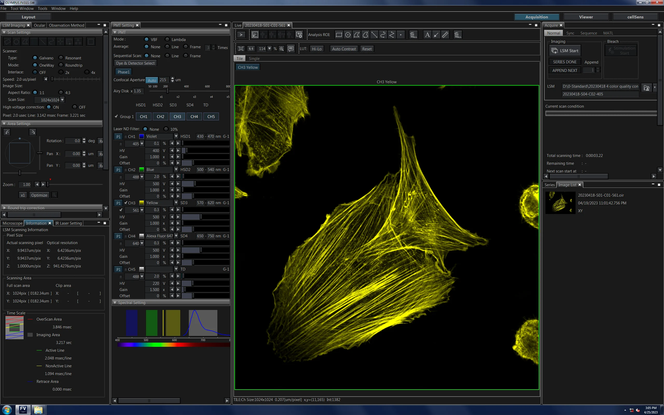
在 2 μs/pixel 的扫描参数下,整张 1024 × 1024 像素图采集耗时约 3.2 s。该参数兼顾了样品的光漂白控制与微管细节的清晰度,是后续多轮检测迭代的基础设置。
20230601Actin-Alexa532 样品检测详情
标记原料
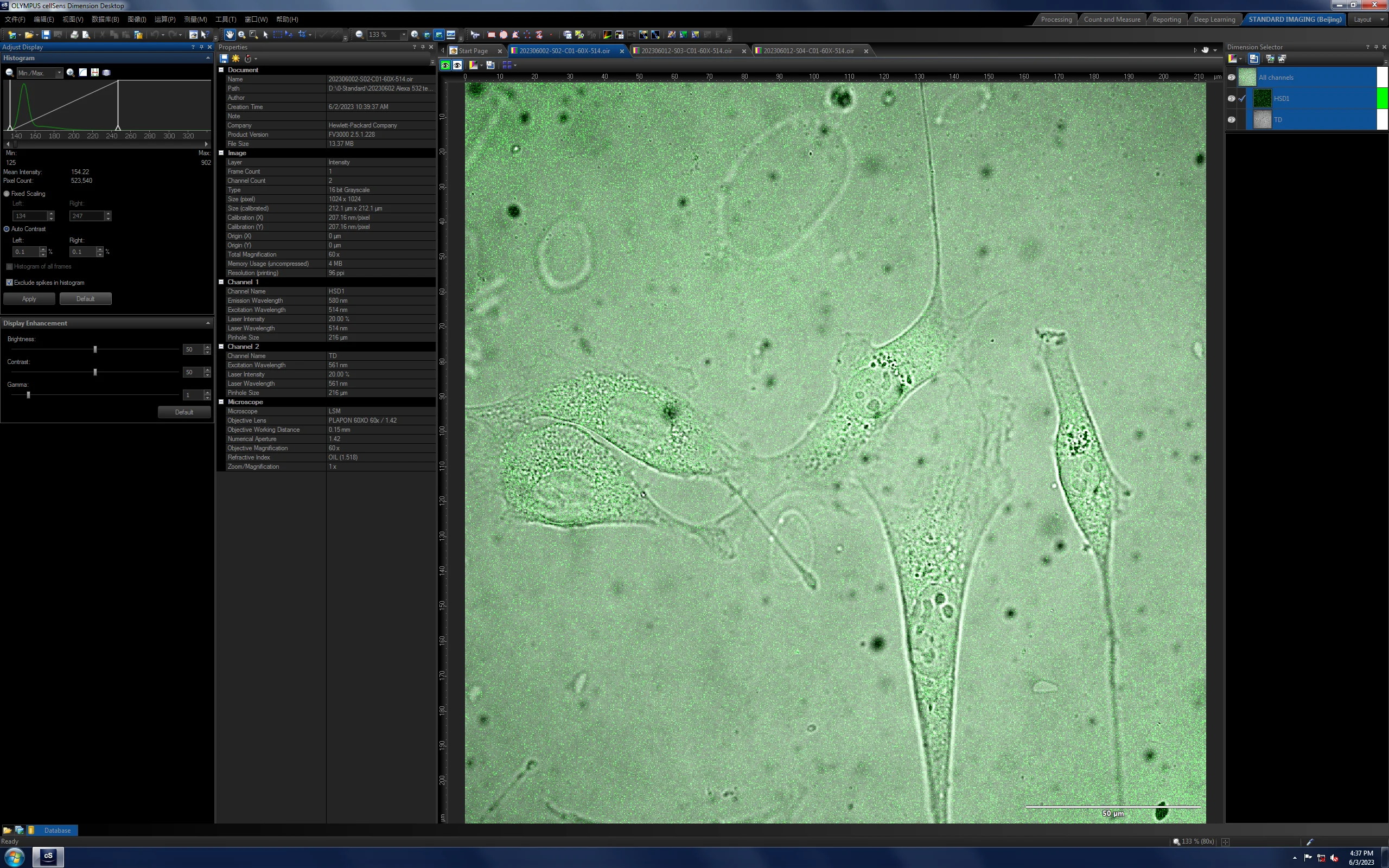
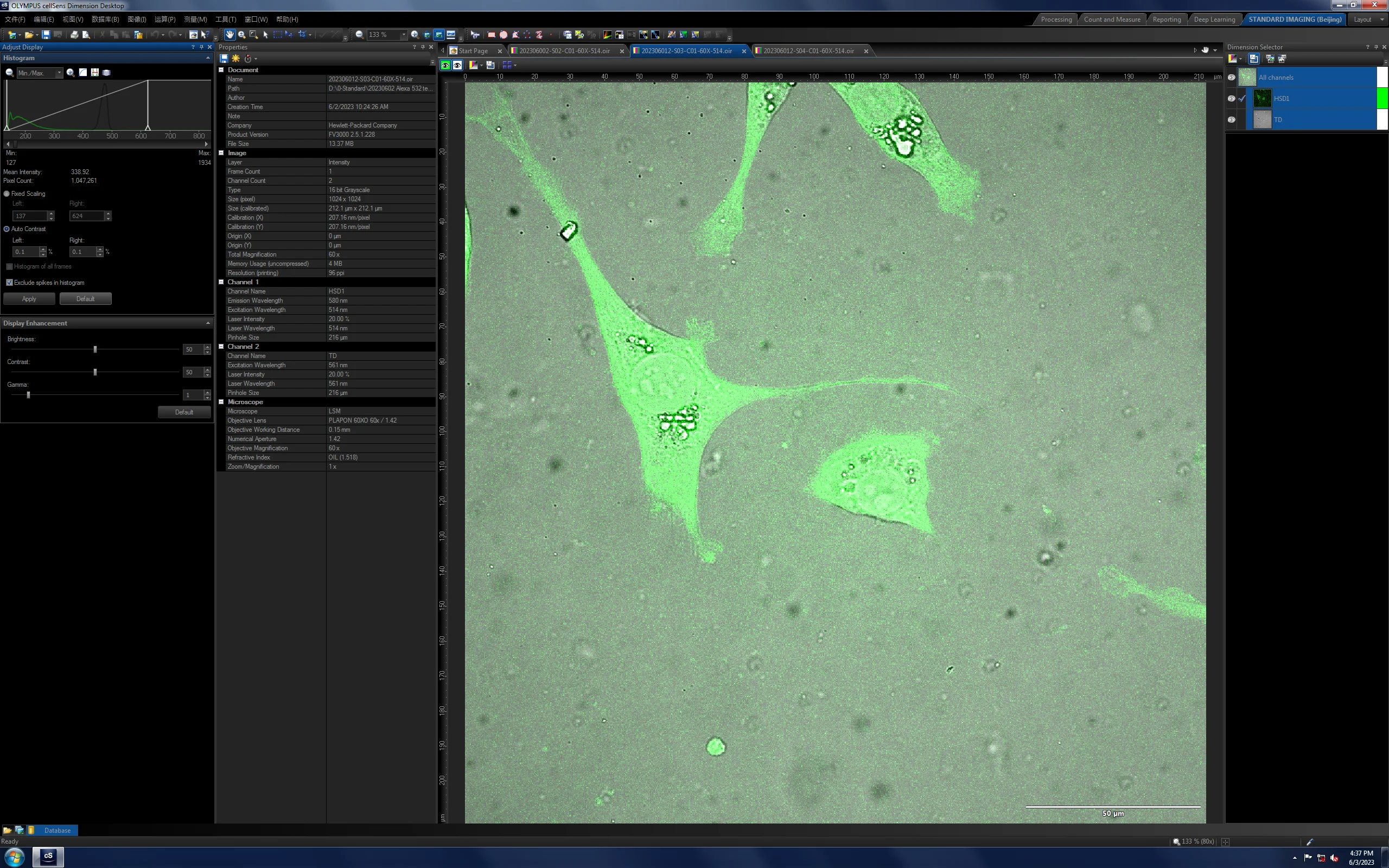
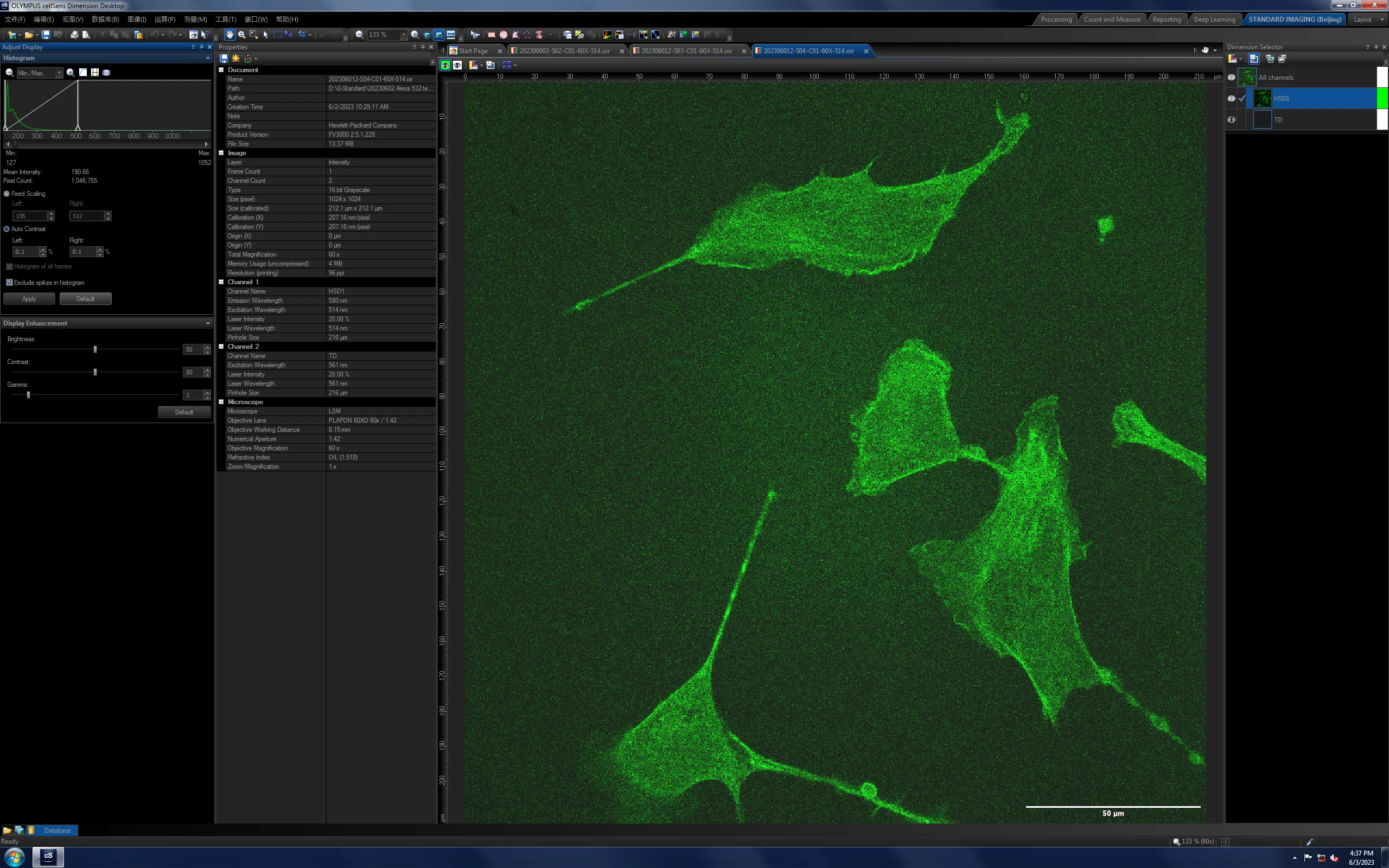
成像检测结果(FV3000 60×)
| 样品 | S01 | S02 | S03 | S04 |
|---|---|---|---|---|
| 染料 | Alexa Fluor 532 phalloidin | Alexa Fluor 532 phalloidin | Alexa Fluor 532 phalloidin | Alexa Fluor 532 phalloidin |
| 浓度 | 4% | 4% | 50% | 16.6% |
| 孵育 | 4 ℃ 过夜(0602) | 4 ℃ 过夜(0602) | 4 ℃ 过夜(0602) | 4 ℃ 过夜(0602) |
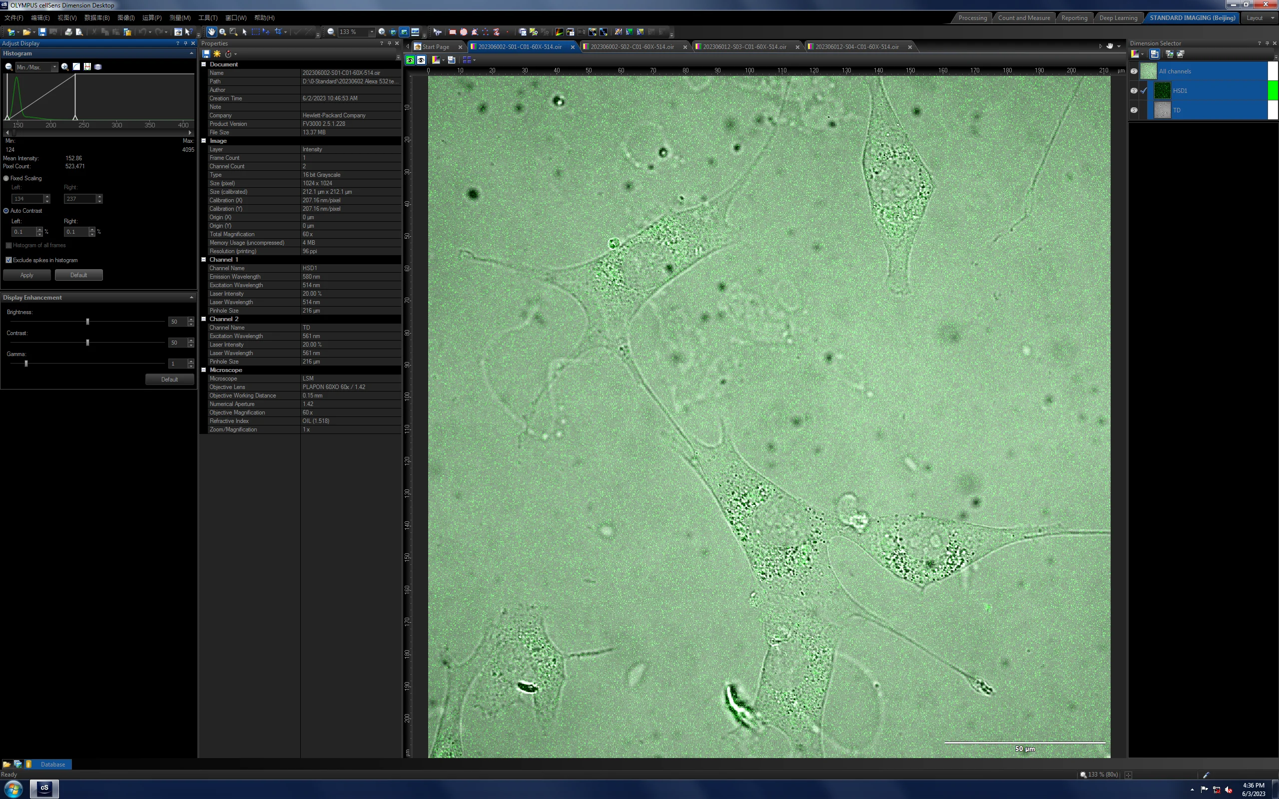
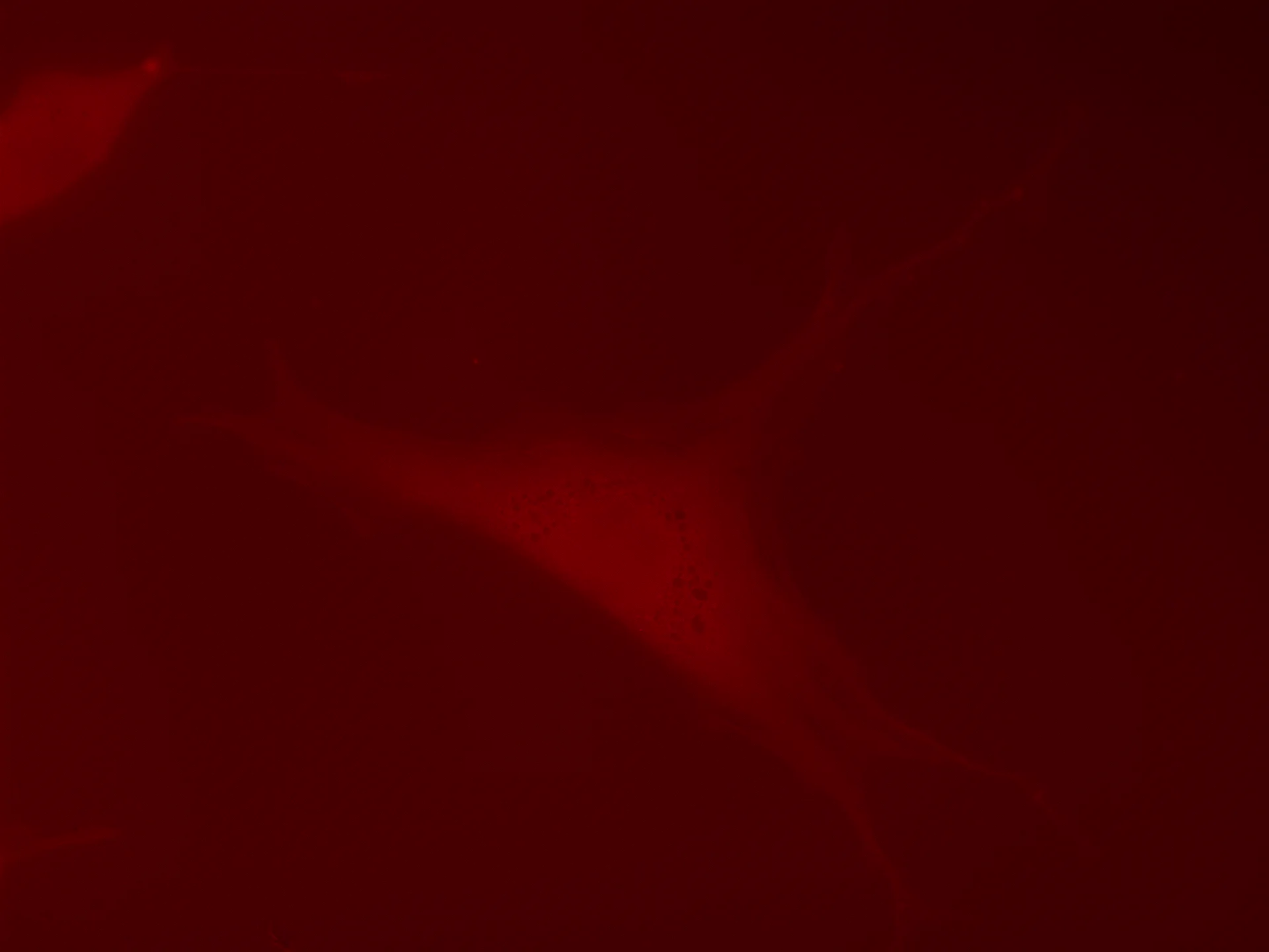
基恩士对照(60×)


20230602Actin-Alexa532 样品检测详情
标记原料
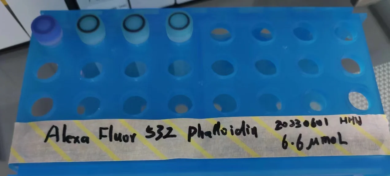
成像检测结果
| 样品 | S01 | S02 | S03 | S04 | S05(+ 标准样) |
|---|---|---|---|---|---|
| 染料 | Alexa Fluor 532 | Alexa Fluor 532 | Alexa Fluor 532 | Alexa Fluor 532 | Alexa Fluor 532 |
| 浓度 | 10% | 50% | 20% | 100% | — |
| 染色时间 | 20230602 | 20230602 | 20230602 | 20230602 | 20230602 |
基恩士初扫(Brightness:1 / 6s)
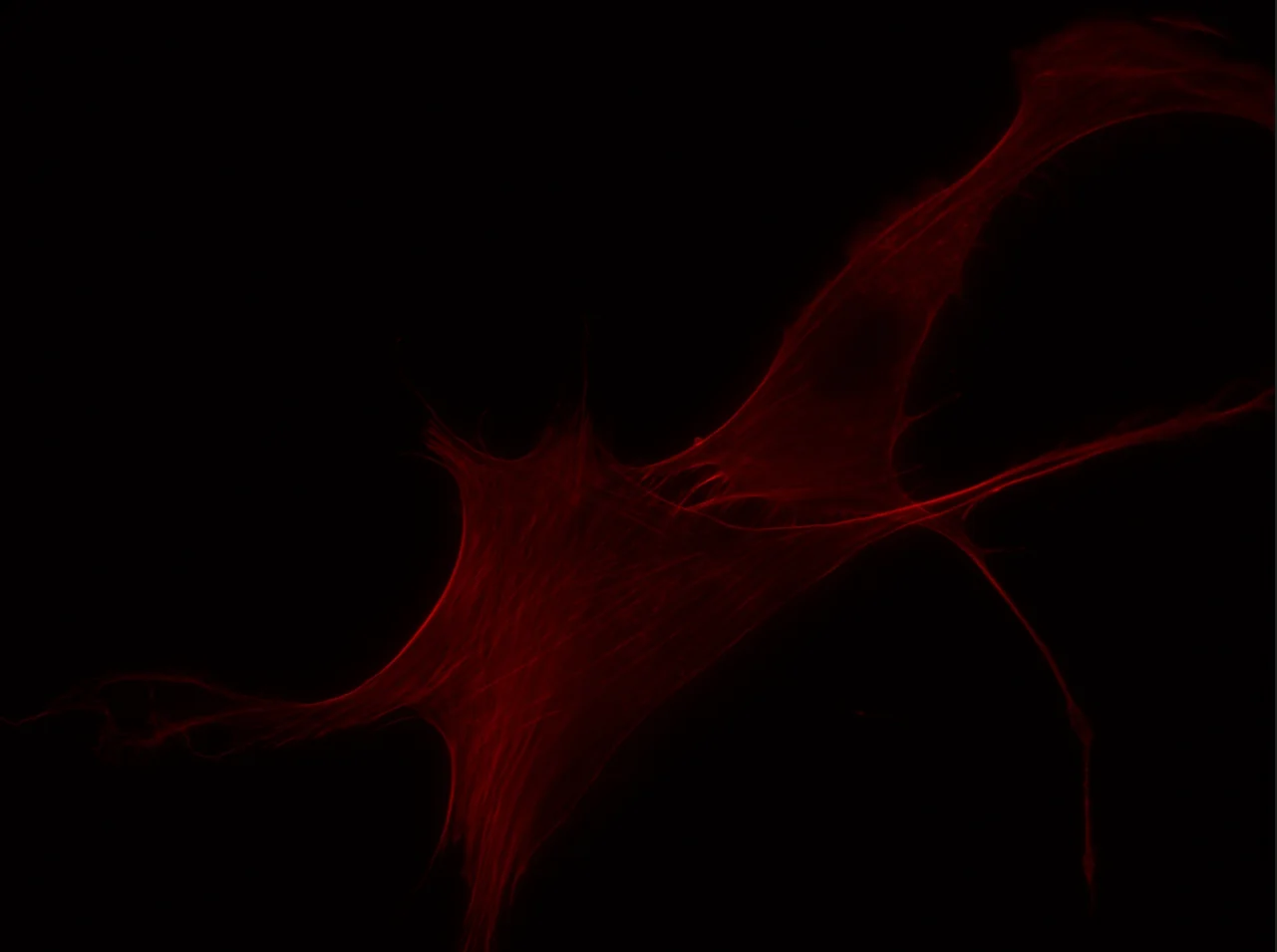
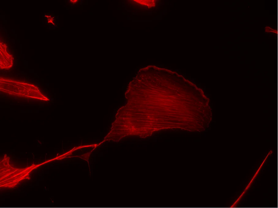
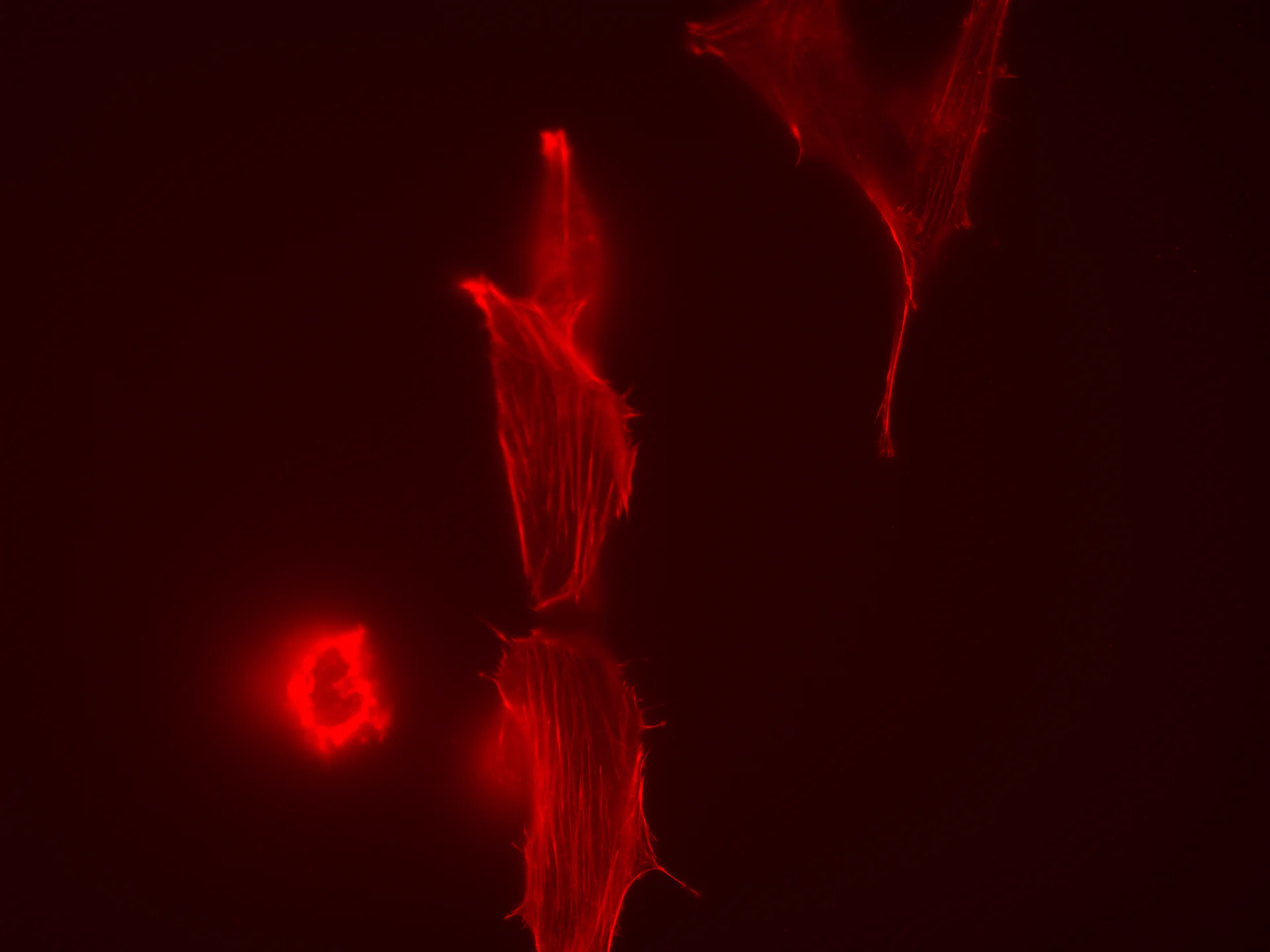
基恩士复测(Brightness:1 / 12s · 4 ℃ 过夜孵育)
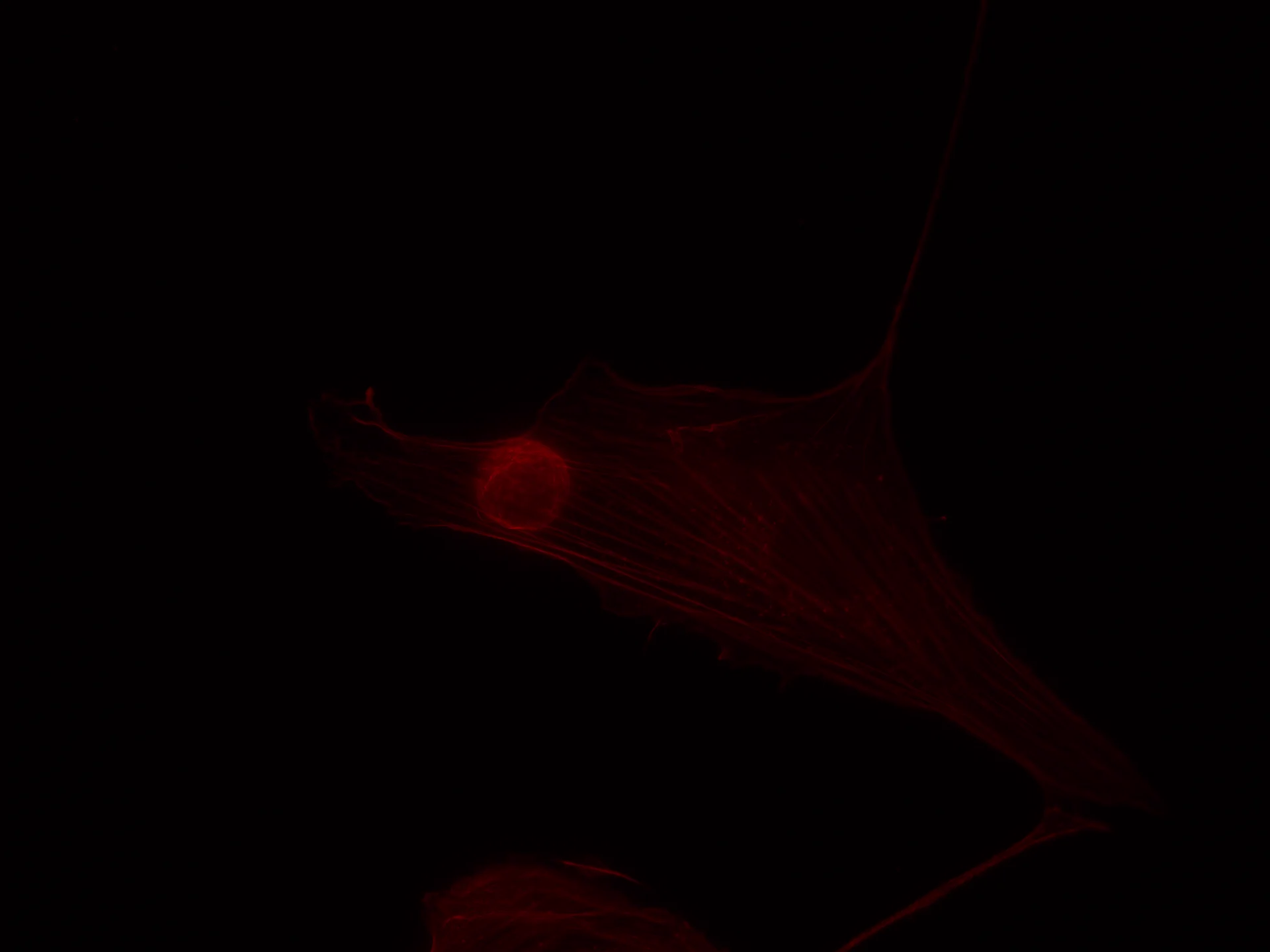
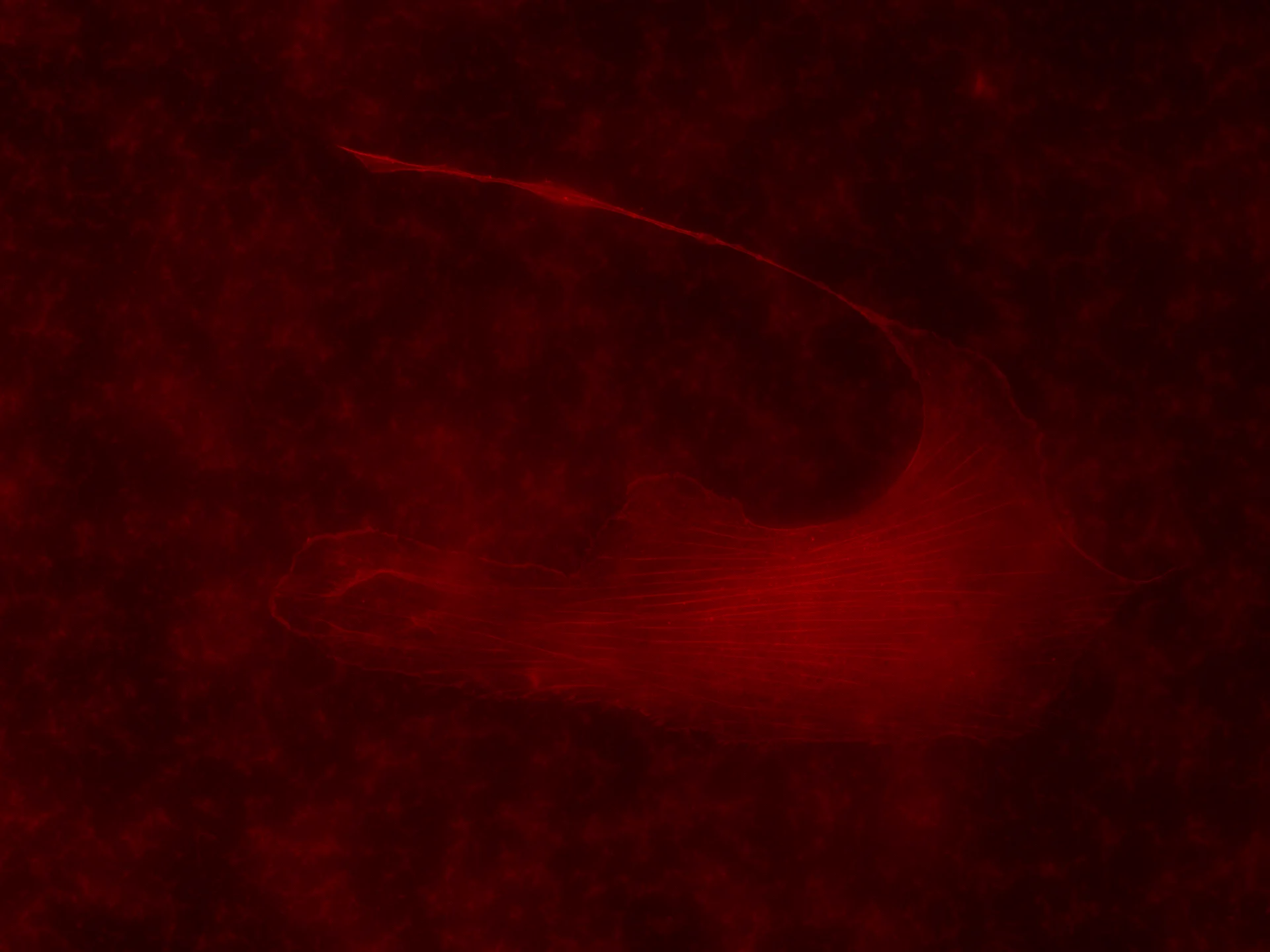
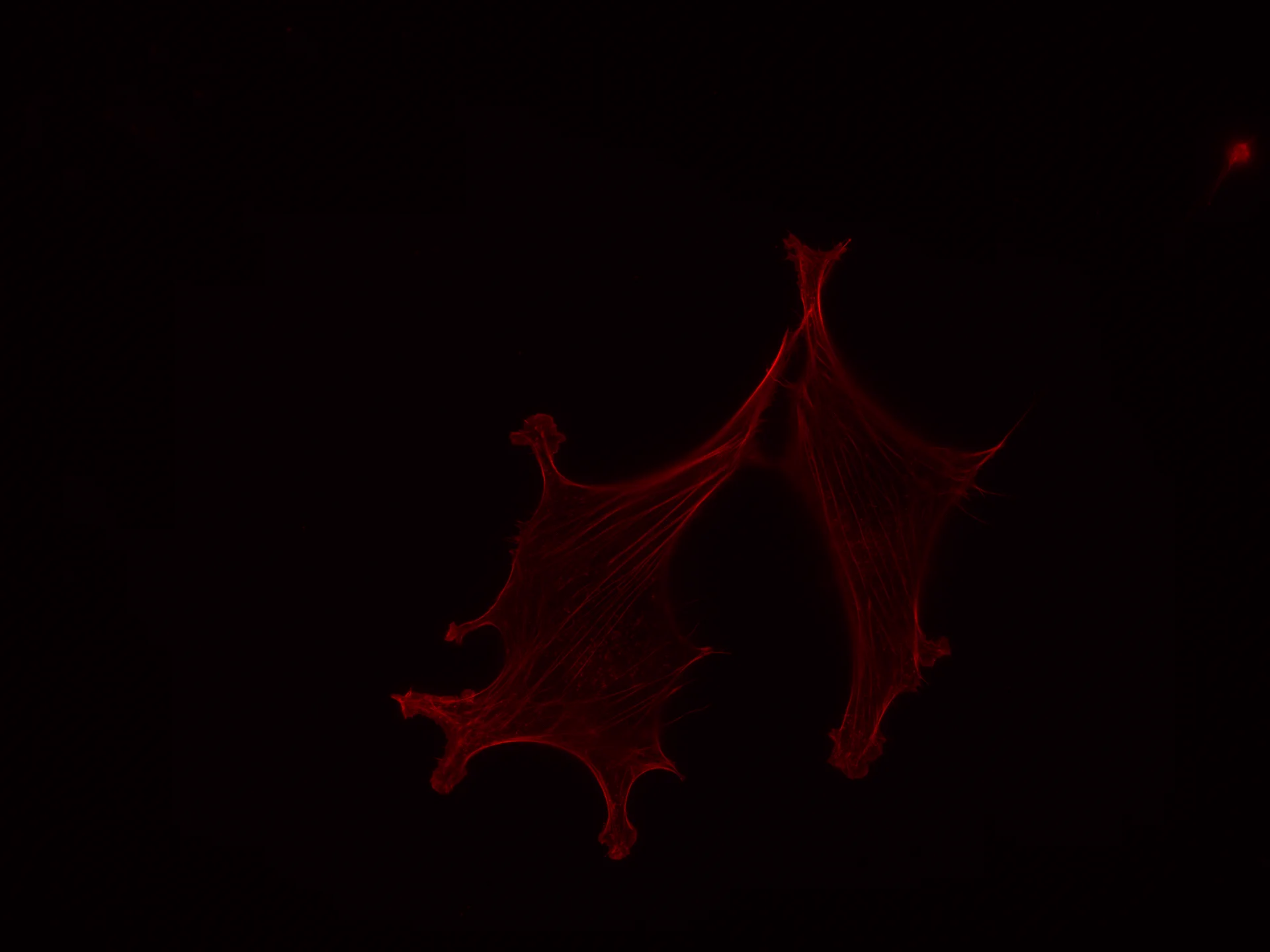
正对照样品(S05)
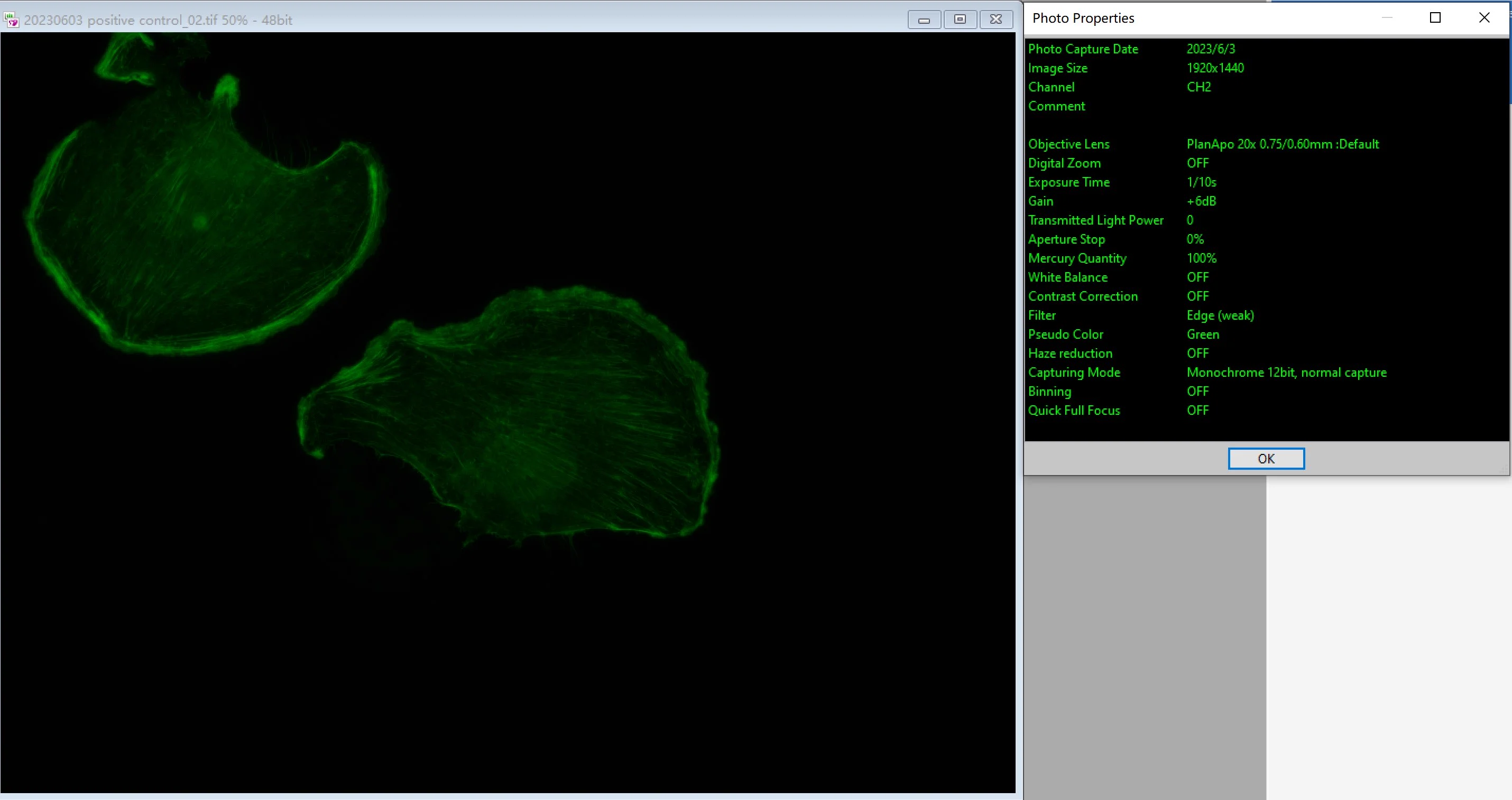
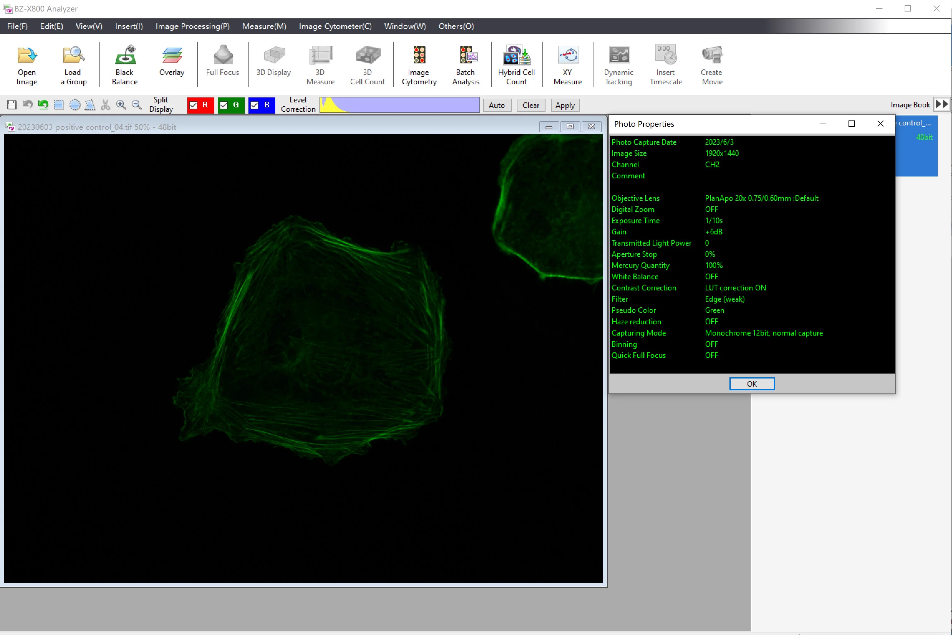
| Positive control(基恩士) | |
|---|---|
| 曝光时间 | 1 / 10 s |
| 通道 | CH2 |
| Gain | +6 dB |
| 激光强度 | 100% |
20230605Actin-Alexa532 浓度优化 · 关键结论
原料记录
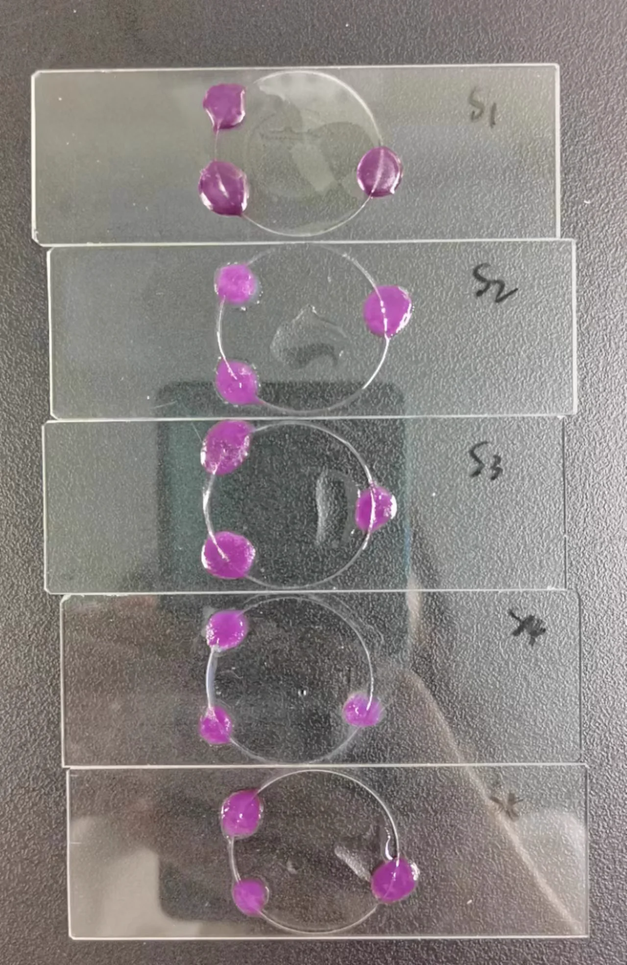
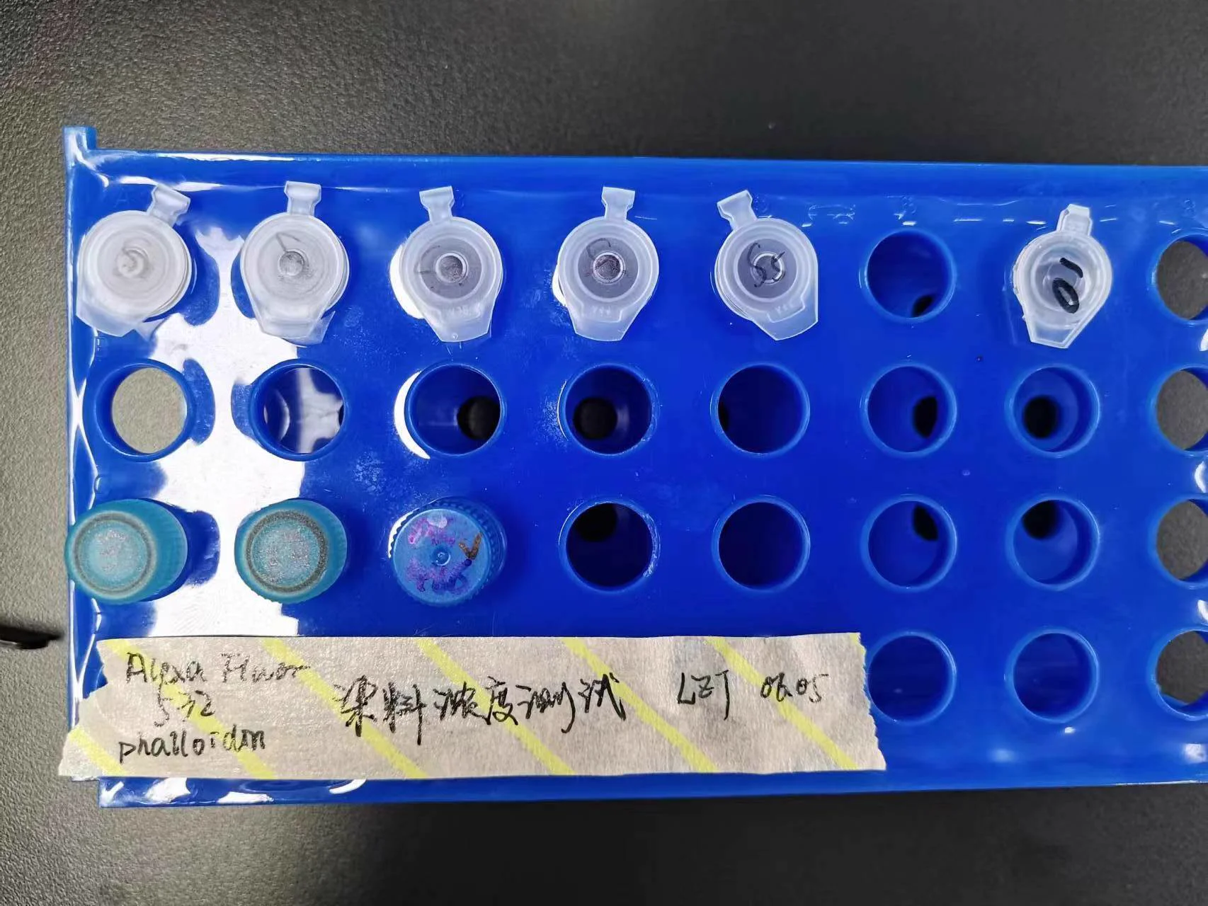
实验条件:环境温度 27 ℃ · 染色时间 3 h
| 样品 | S01 | S02 | S03 ⭐ 最优 | S04 | S05 正对照 |
|---|---|---|---|---|---|
| 染料浓度 | 50% | 100% | 200% | 300% | 488 标准 |
| 配置体系 | 染料(甲醇)12.5 μL + Buffer 12.5 μL |
染料(DMSO)2.5 μL + Buffer 22.5 μL |
染料(DMSO)5 μL + Buffer 20 μL |
染料(DMSO)7.5 μL + Buffer 17.5 μL |
488 1 μL + Buffer 24 μL |
20× 时序 cycle
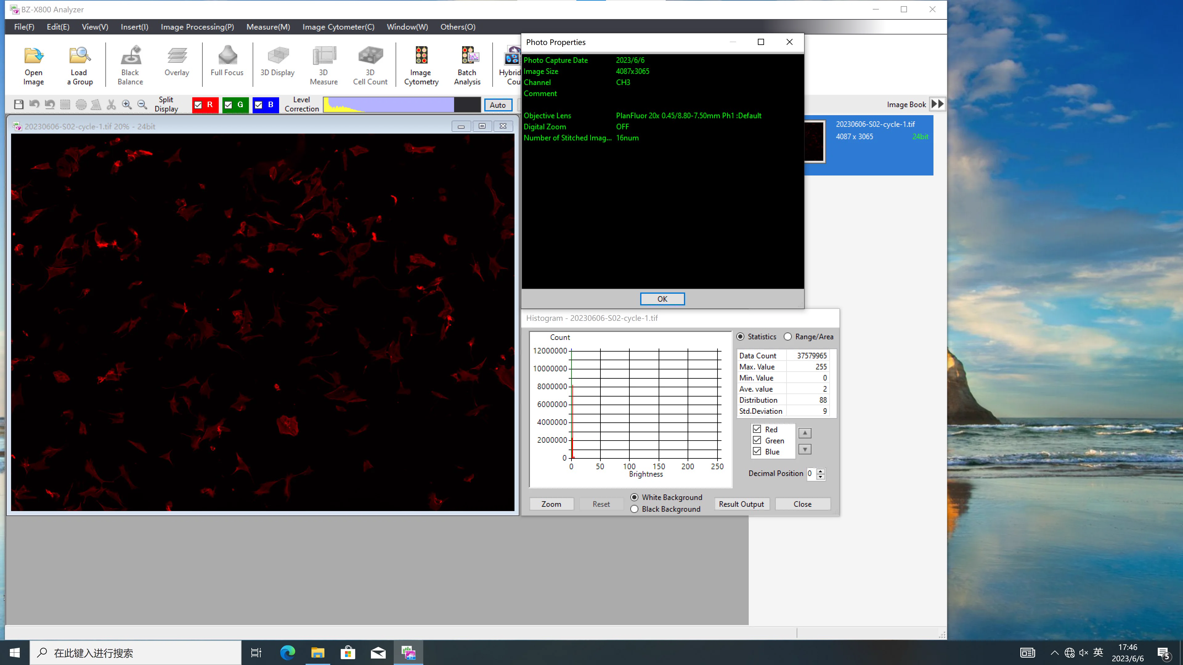
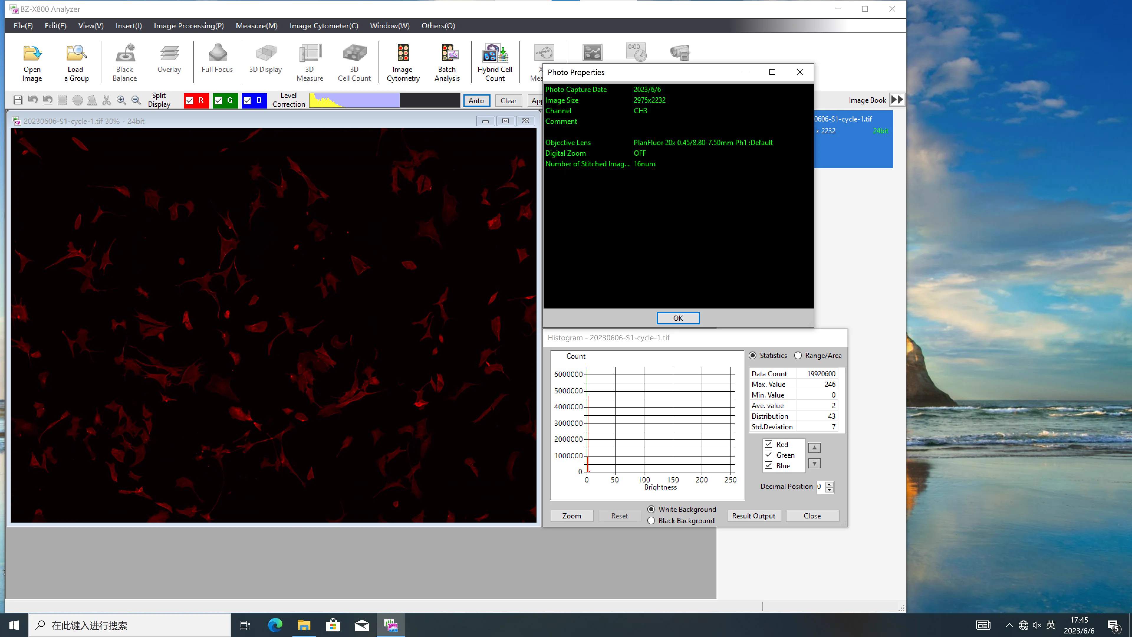
60× 共聚焦单帧(6.5 日)
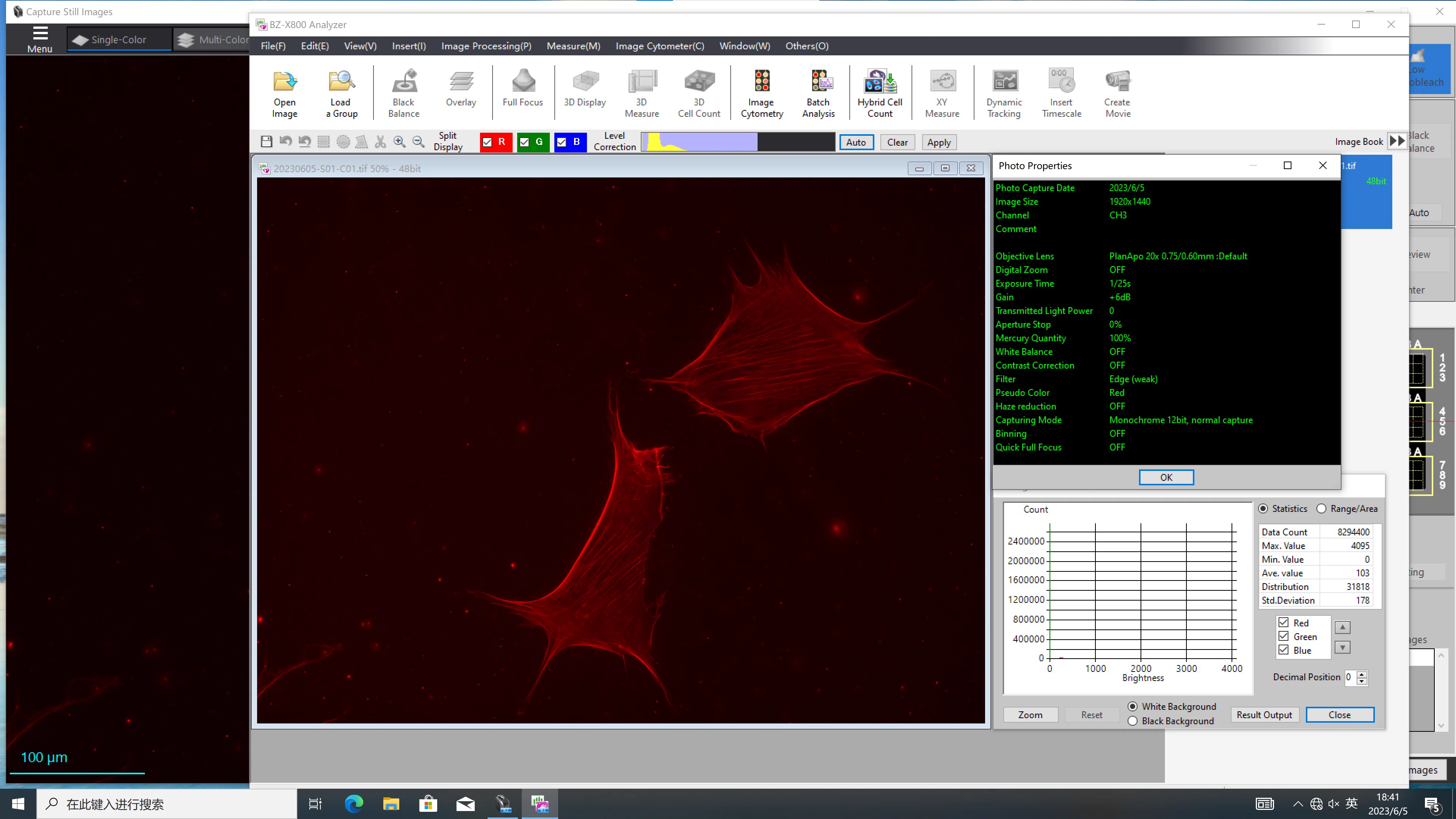
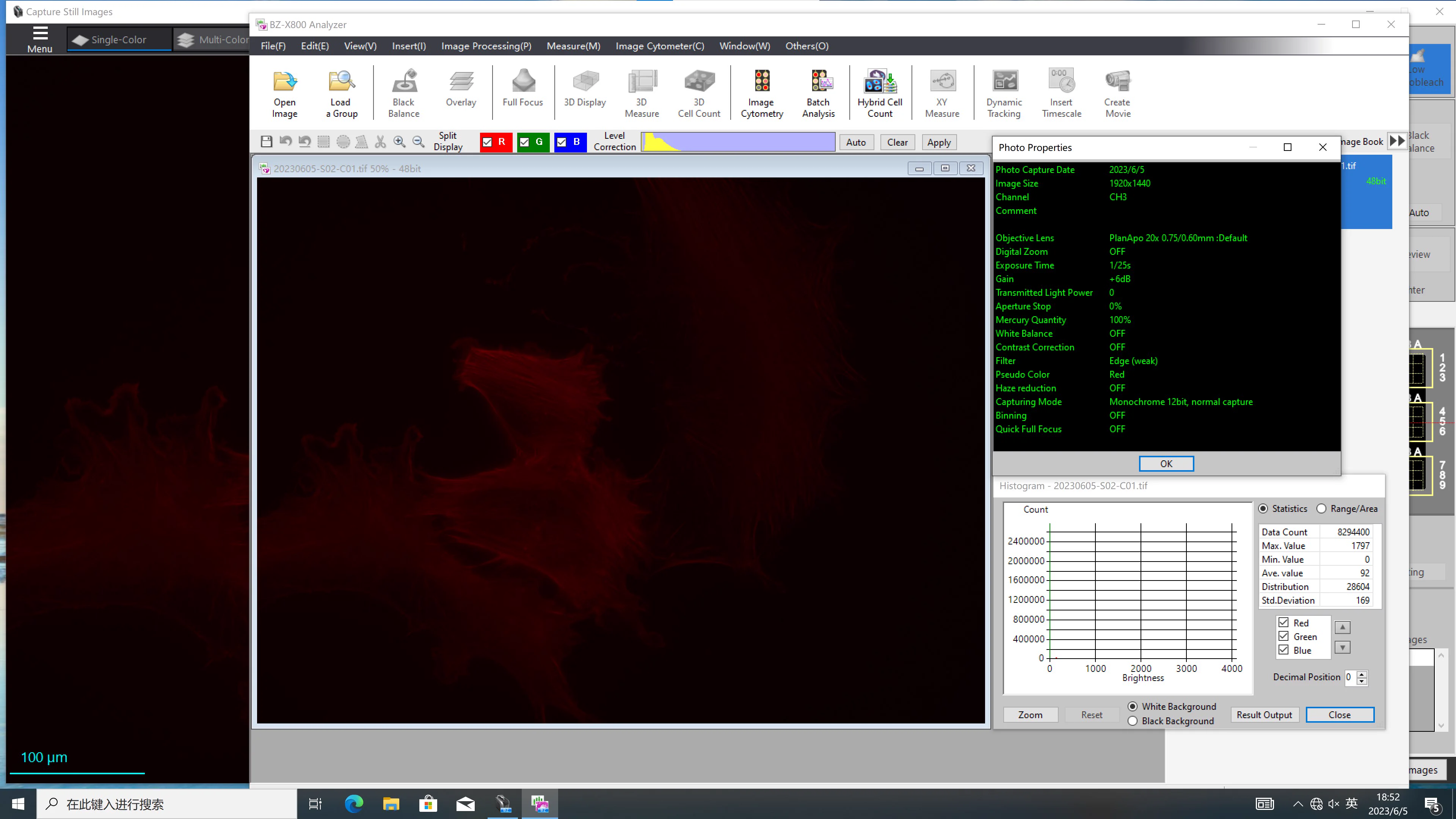
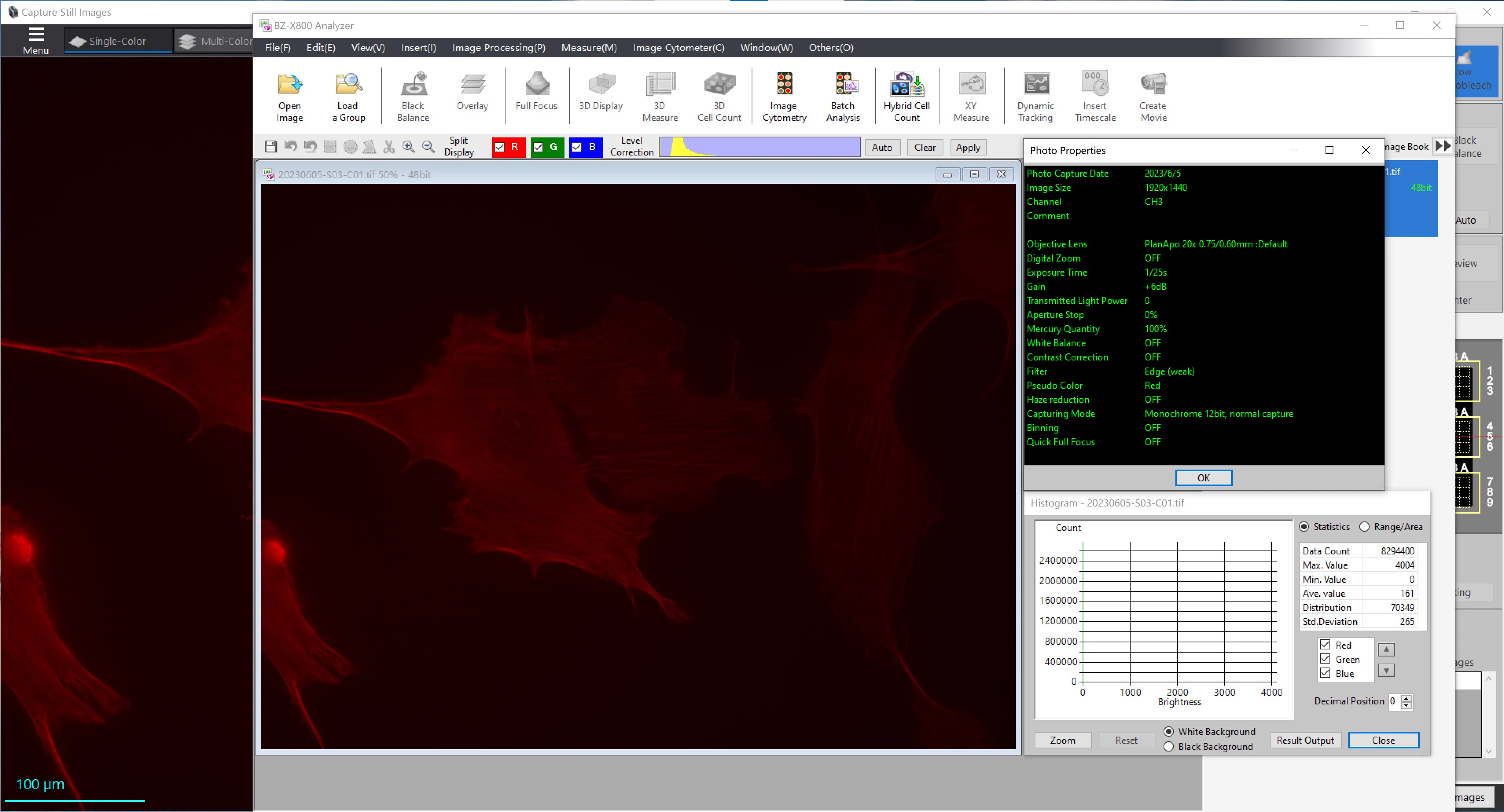
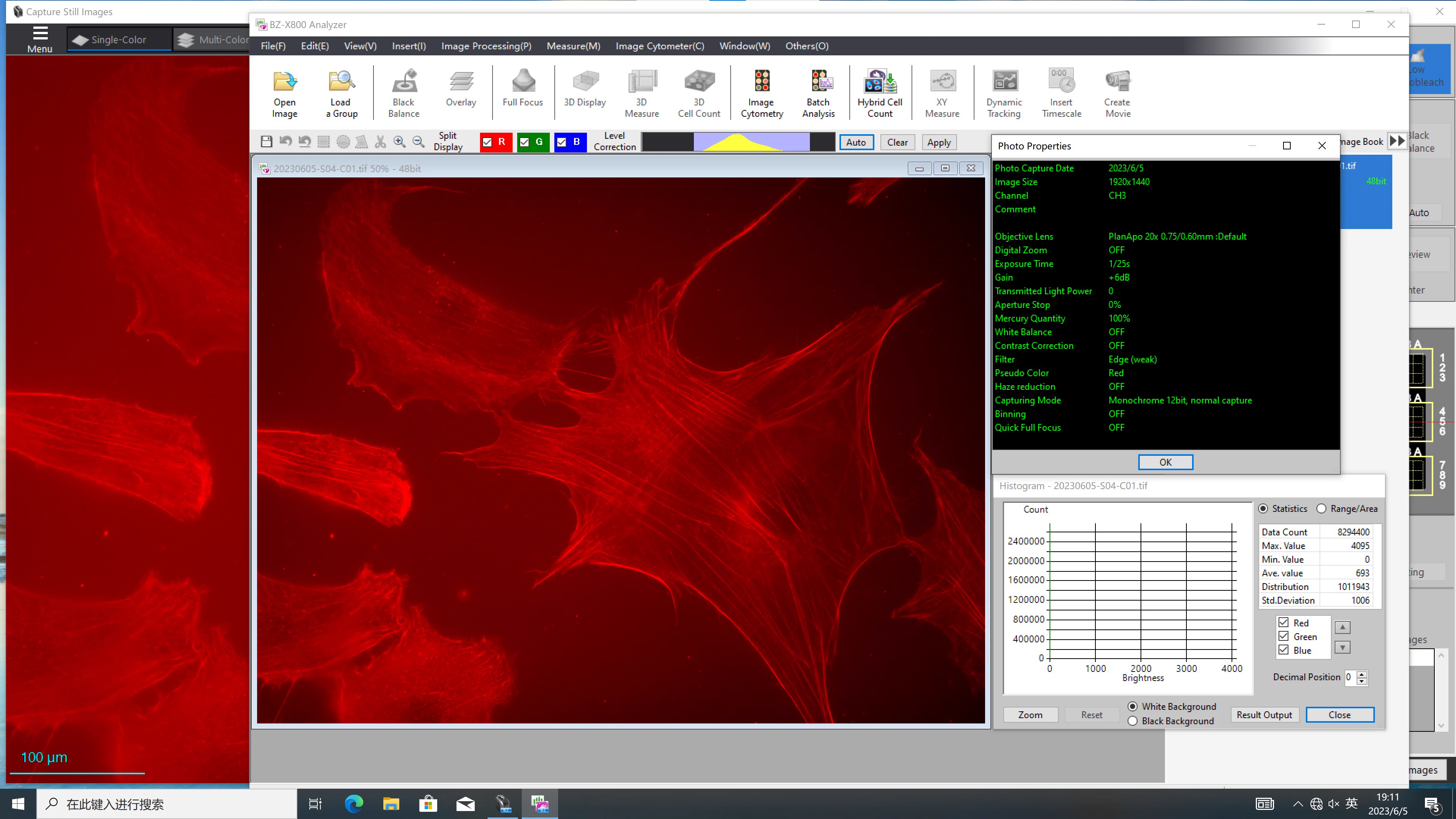
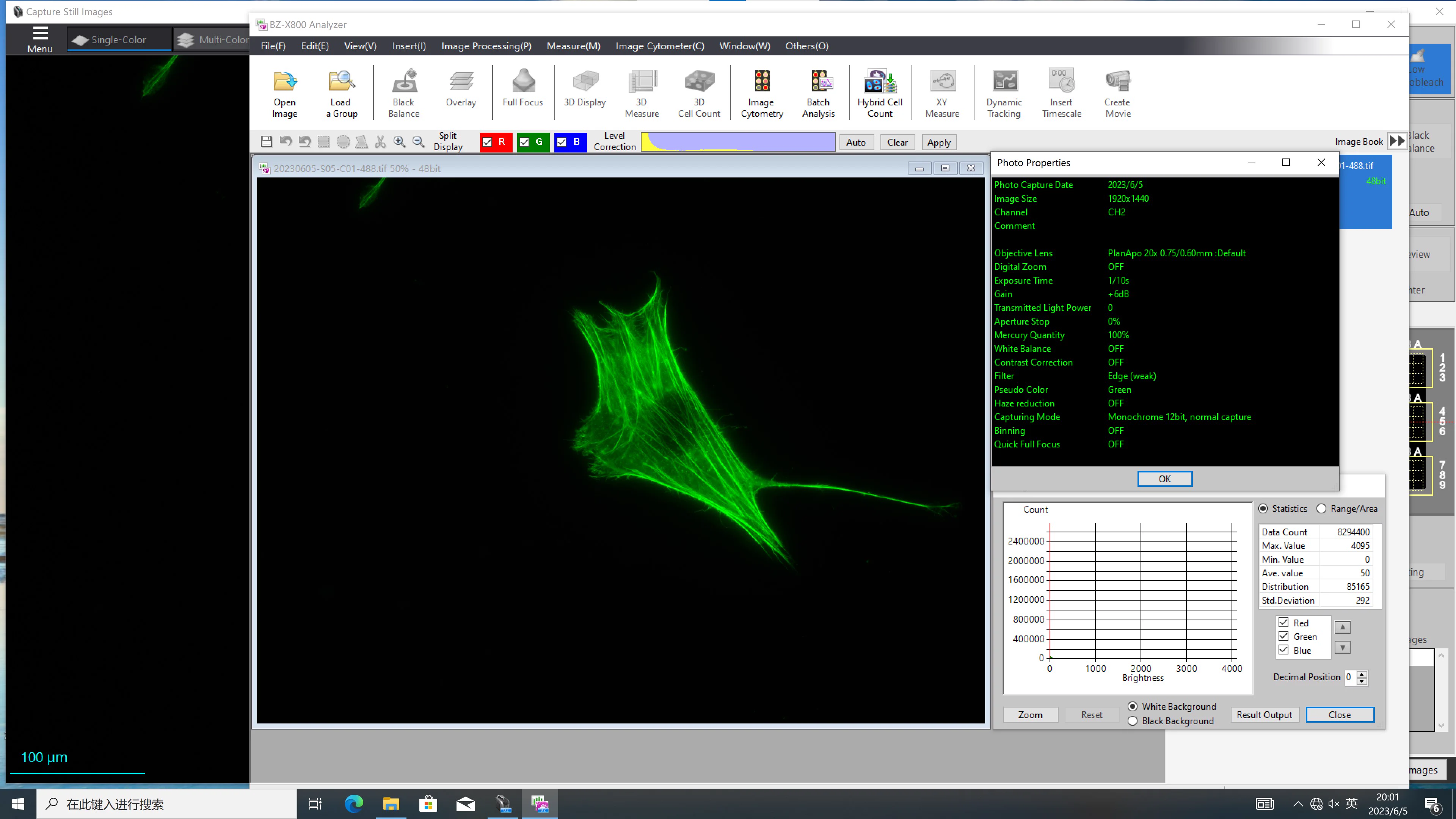
60× 532 复测(6.6 日,S03/S04)
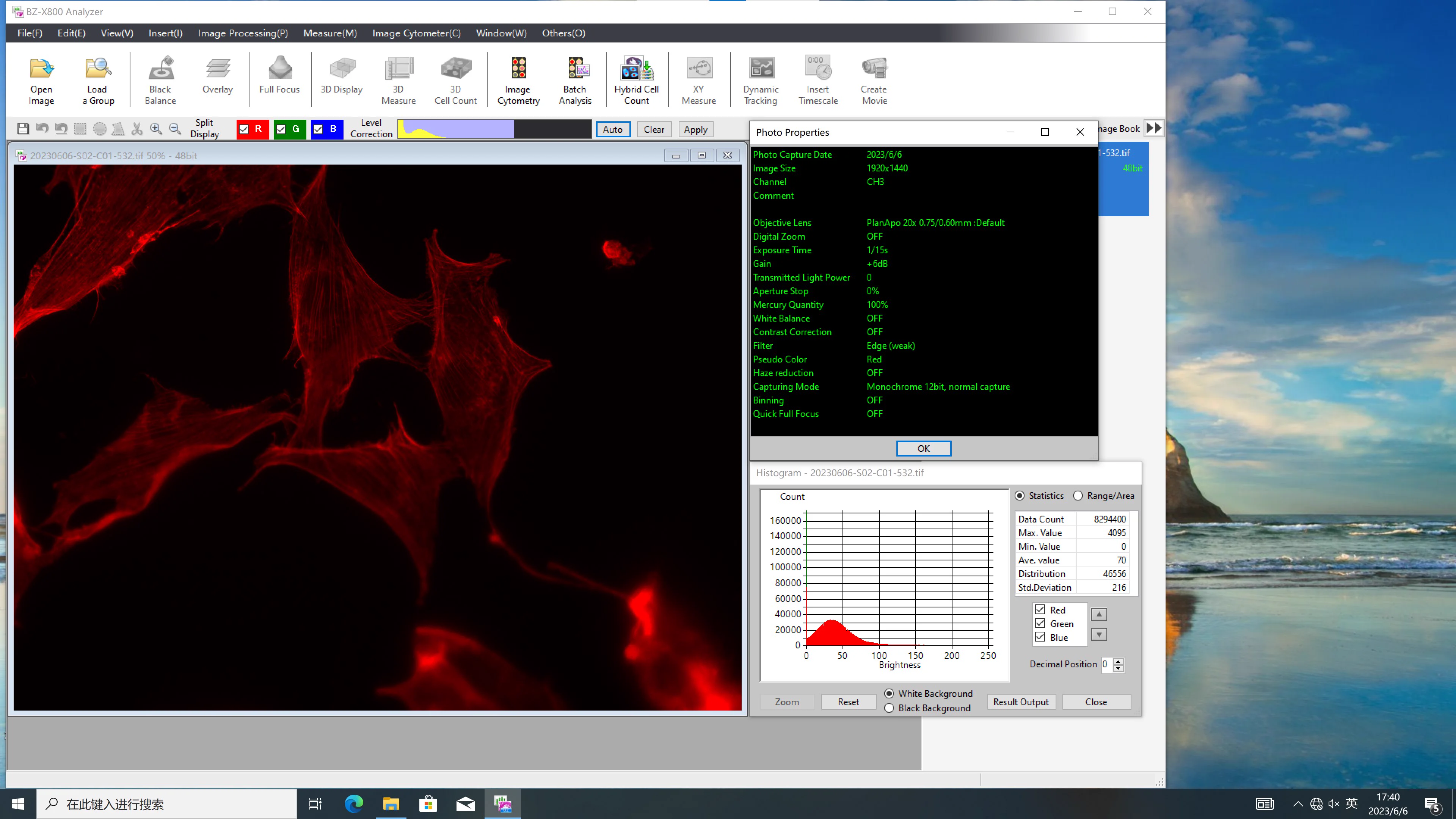
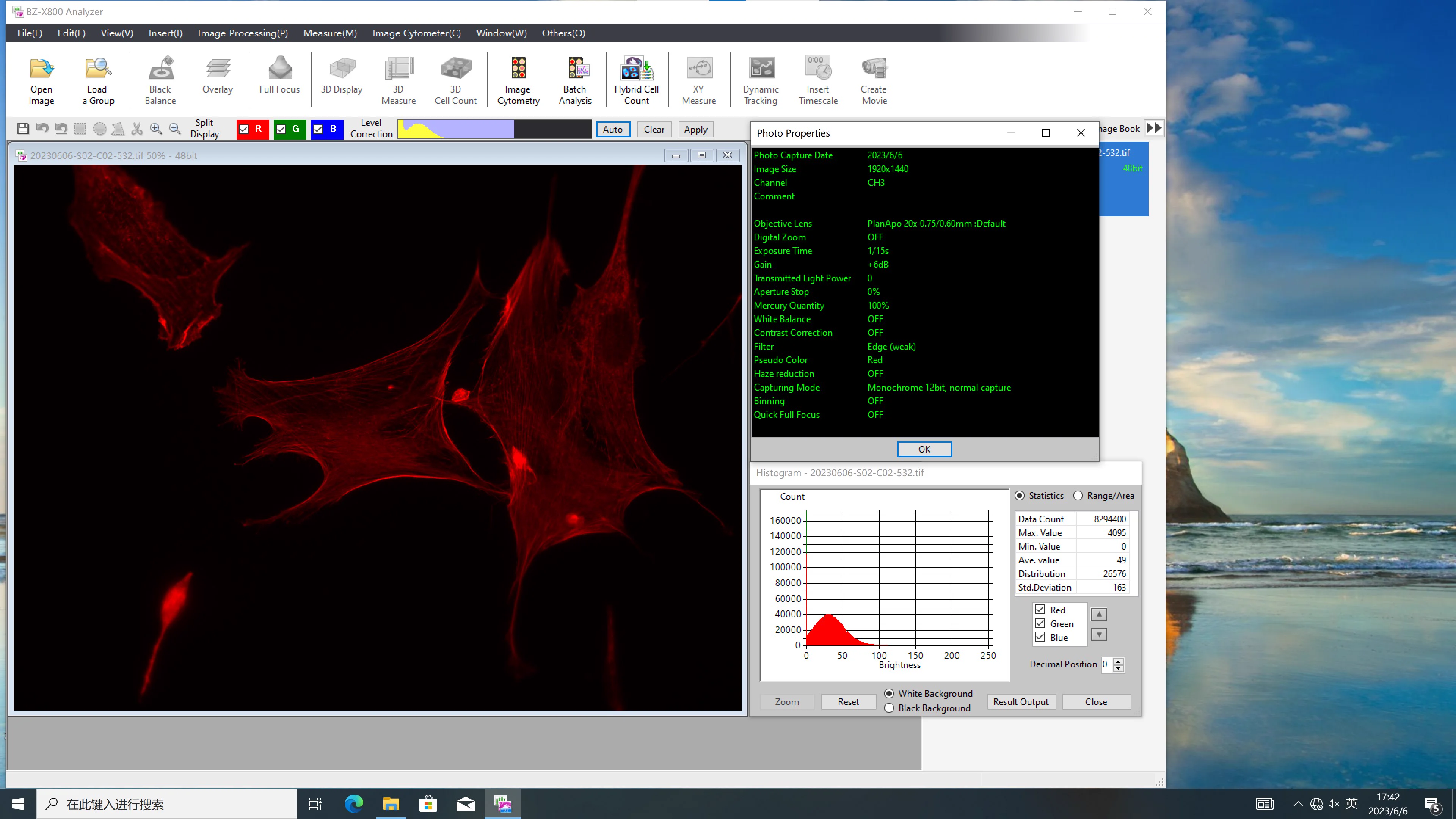
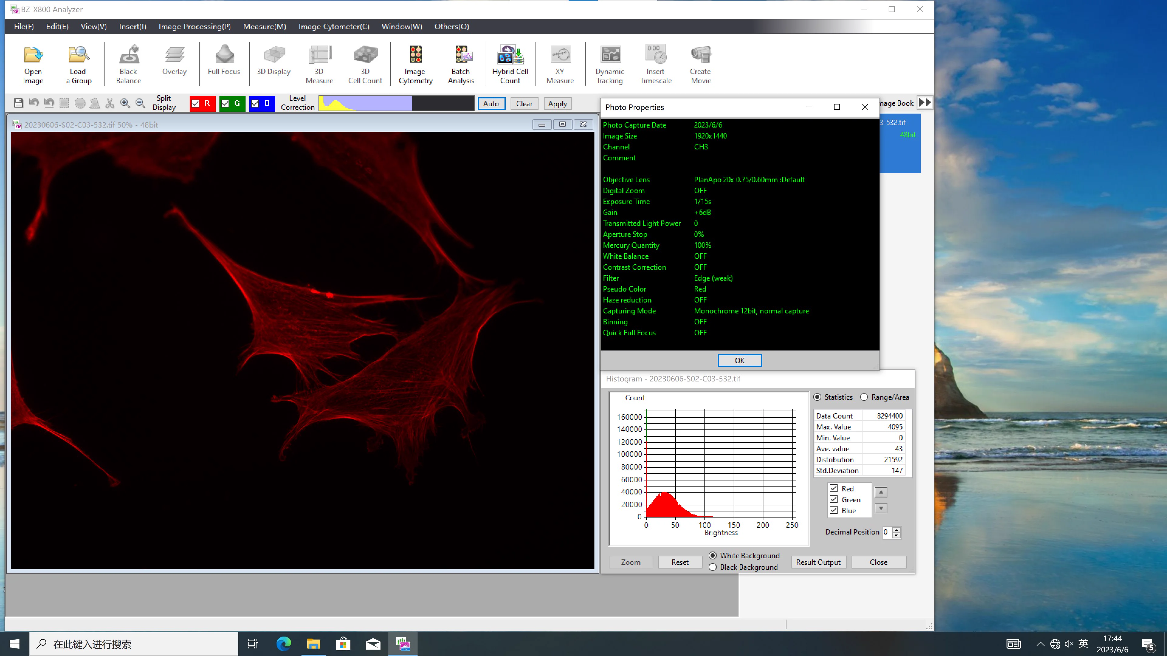
从染料自身看,该批次 Alexa 532 染料质量较差,需 50× 于正对照染料浓度才能达到相同效果,建议更换染料。
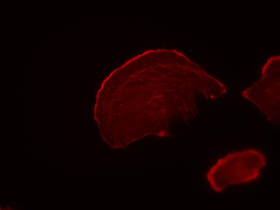
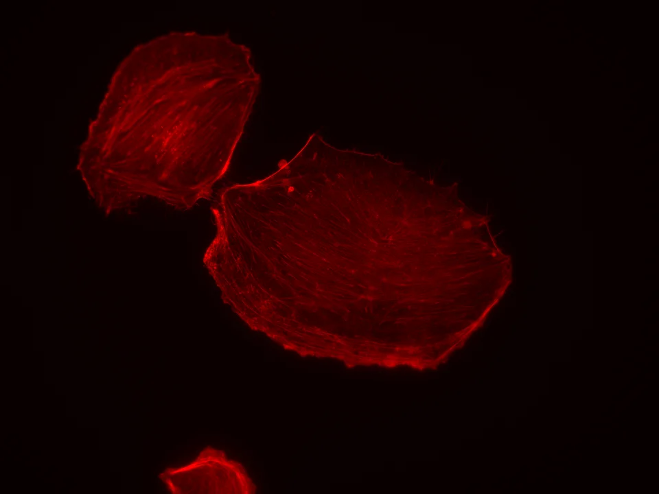
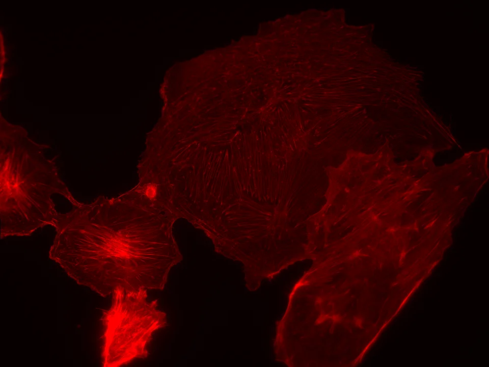
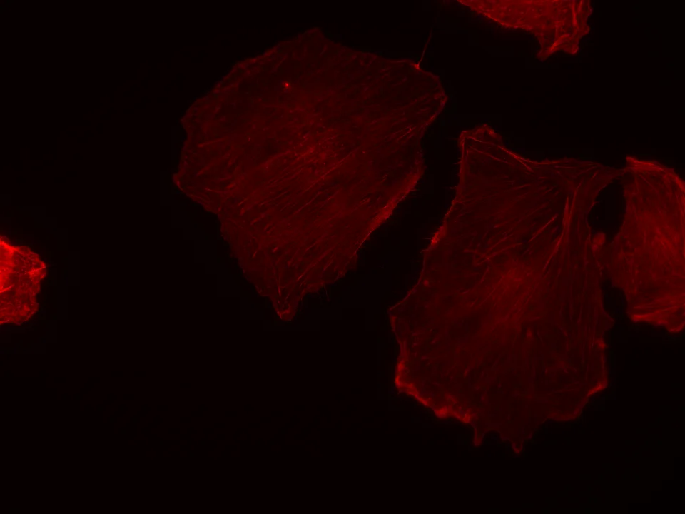
20230614Actin-Alexa532 样品检测报告
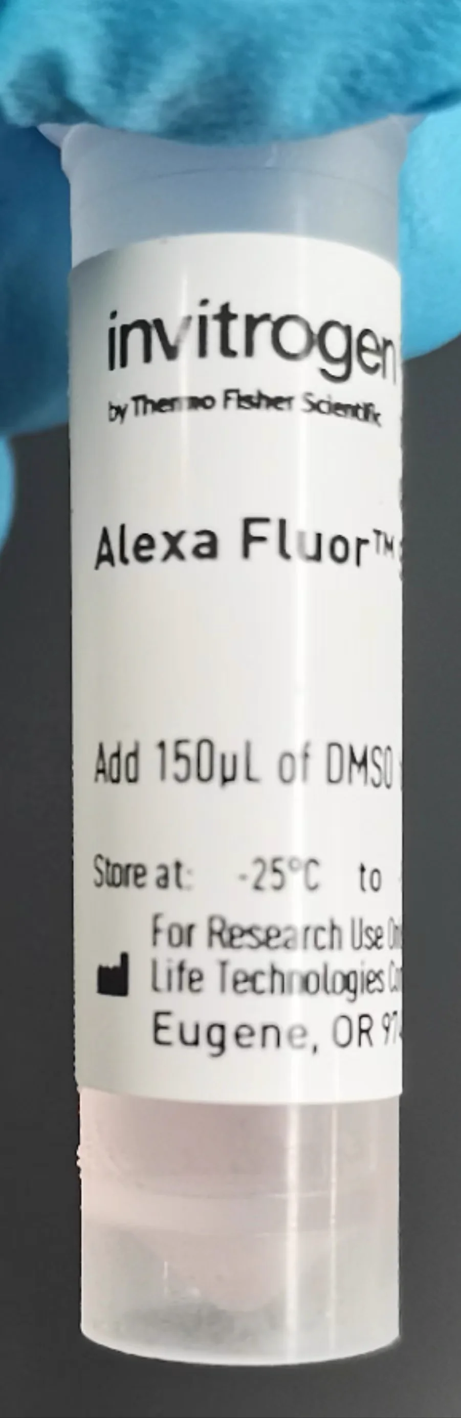
标记原料
成像结果(S1 · 60× 532 + 明场 BF)
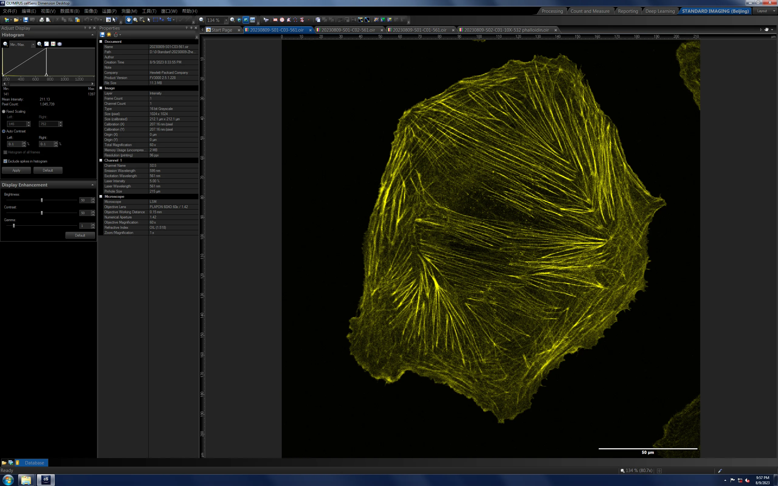
20230809Actin-Alexa532 样品检测详情
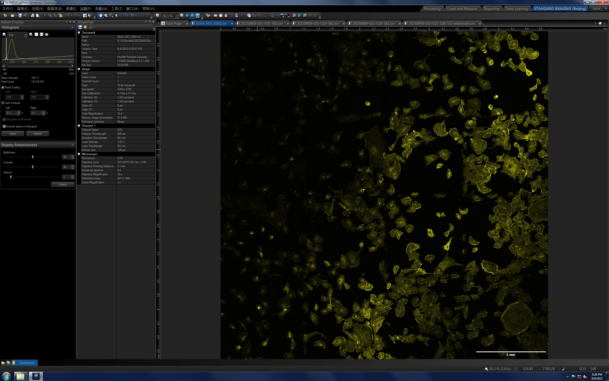
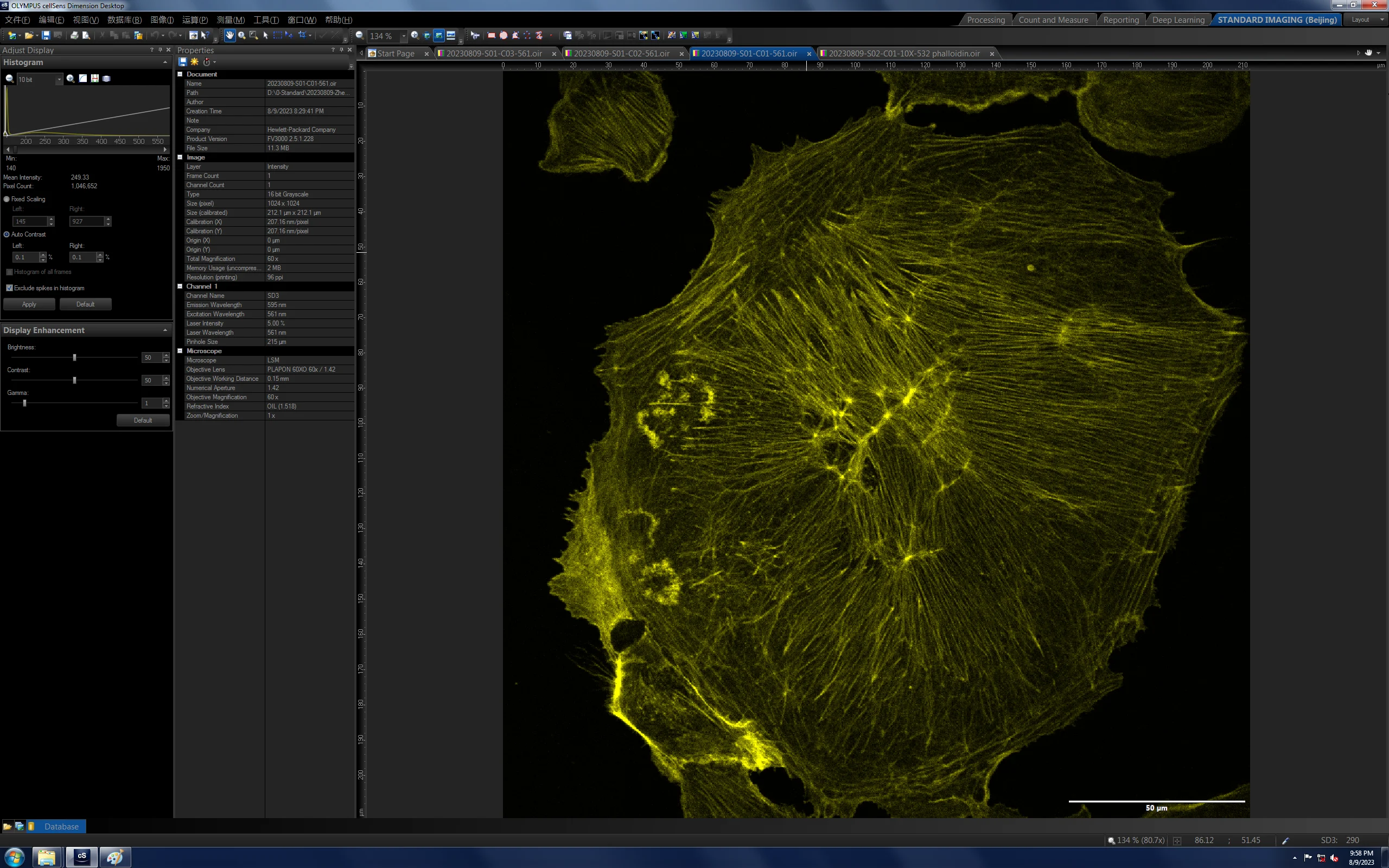
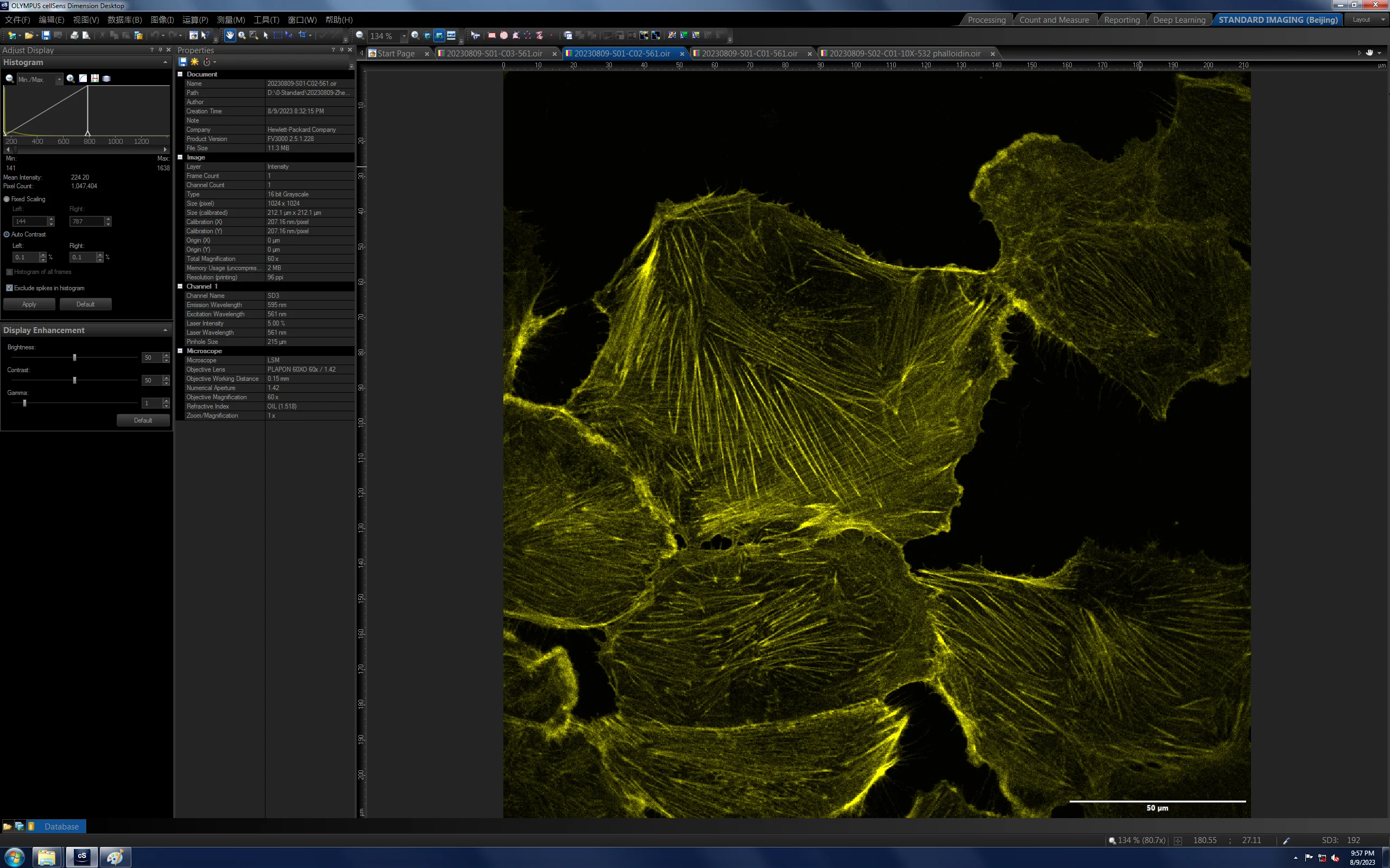
60× 532 成像
Protocol实验流程
🧪细胞爬片表面处理
- 将洁净的细胞爬片放入六孔板或 3.5 cm 培养皿中,加入 1–2 mL 完全培养基使之浸润爬片;
- 放入培养箱孵育 12 h 以上,使用前吸除培养基。
🧫成像细胞接种
- 取细胞状态最优的培养皿消化处理,消化离心后细胞计数,确保每孔(六孔板)/ 每皿 内有 4 × 10⁴ 个活细胞,计算所需细胞总量;
- 取出所需细胞,按每孔/每皿加 2 mL 培养基计算,配制成细胞悬液并加入到孔板或培养皿中;
- 成像细胞培养需 1–2 天,观测到细胞状态良好且单个细胞舒展不拥挤、面积大、细胞透光性强透明度高时,可进行后续成像步骤。
🎨染色步骤
- 细胞固定:使用 4% PFA 固定细胞形态;
- 背景荧光淬灭:使用硼氢化钠消除背景荧光;
- 细胞封闭:使用 BSA 处理细胞,使其结合位点更易结合染料;
- 细胞染色:使用 Alexa 532 染料染色细胞,过夜孵育;
- 细胞后固定:再次使用 PFA 固定细胞形态,使样片形态更加稳定。
⚠️ 注意:每一步细胞处理后都需要对细胞进行清洗,去除上一步使用的试剂。
Methods染色过程描述
🧬Actin staining(肌动蛋白染色)
Cell culture
The Biologics Standards-Cercopithecus-1 (BSC-1) cell line and NIH/3T3 cell line were purchased from Pricella Life Technology Co. LTD. BSC-1 cells and NIH/3T3 cells were cultured in DMEM (Invitrogen, #11965-118) supplemented with 10% fetal bovine serum (FBS) (Gibco, #16010-159). To prevent bacterial contamination, 100 μg/mL penicillin and streptomycin (Invitrogen, #15140122) were added in the DMEM medium. Cells were grown under standard cell culture conditions (5% CO₂, humidified atmosphere at 37 ℃). BSC-1 cells were plated on #1.5 glass-bottom dish over 48 h before sample preparation. For cell passage, cells were washed with pre-warmed PBS (Life Technologies, #14190500BT) 3 times and digested with 25% trypsin (Gibco, #25200-056) for 30 s. BSC-1 cell line were tested for potential mycoplasma contamination (MycoAlert, Lonza), and all tests showed negative results.
Cell staining
Cells were grown on 35 mm, #1.5 glass coverslips (Standard Imaging (Beijing) Biotech, STGBD-035-1). To increase cell adhesion, we pre-treated glass-bottom dishes with fibronectin (Invitrogen, #33016015) for 1 h at 37 ℃. On the day of sample preparation, the cell density should be about 50%–70%. Cells were fixed with 37 ℃ pre-warmed fixation buffer for 10 min, containing 4% paraformaldehyde (EMS) and 0.1% glutaraldehyde in PBS. Then the sample were washed three times with PBS. For quenching the background fluorescence, we incubate the cells with 2 mL 0.1% NaBH₄ solution in PBS for 7 min, optionally shake on the shaker (<1 Hz). The sample were washed three times with 2 mL PBS and then incubated for 30 min in PBS containing 5% BSA (Jackson, #001-000-162) and 0.5% Triton X-100 (Fisher Scientific) at 37 ℃. All dyes were diluted in the 5% BSA + 0.5% triton solution described above. Next, we incubate the sample for 40 min with the appropriate dilution of dyes: phalloidin-Alexa532 (ThermoFisher, A22282) or Acti-Stain 555 Phalloidin (Cytoskeleton, PHDH1-A) at 25 ℃. After washed 3 times with PBS, cells were fixed with post-fixation buffer for 10 min. The samples were stored in 4 ℃ in PBS no more than one week.
🧬Tubulin staining(微管染色)
Cell culture
The Biologics Standards-Cercopithecus-1 (BSC-1) cell line was purchased from Pricella Life Technology Co. LTD. BSC-1 cells were cultured in DMEM (Invitrogen, #11965-118) supplemented with 10% fetal bovine serum (FBS) (Gibco, #16010-159). To prevent bacterial contamination, 100 μg/mL penicillin and streptomycin (Invitrogen, #15140122) were added in the DMEM medium. Cells were grown under standard cell culture conditions (5% CO₂, humidified atmosphere at 37 ℃). BSC-1 cells were plated on #1.5 glass-bottom dish over 48 h before sample preparation. For cell passage, cells were washed with pre-warmed PBS (Life Technologies, #14190500BT) 3 times and digested with 25% trypsin (Gibco, #25200-056) for 30 s. BSC-1 cell line were tested for potential mycoplasma contamination (MycoAlert, Lonza), and all tests showed negative results.
Immunofluorescence staining
Cells were grown on 35 mm, #1.5 glass coverslips (Standard Imaging (Beijing) Biotech, STGBD-035-1). To increase cell adhesion, we pre-treated glass-bottom dishes with fibronectin (Invitrogen, #33016015) for 1 h at 37 ℃. On the day of sample preparation, the cell density should be about 50%–70%. Cells were fixed with 37 ℃ pre-warmed fixation buffer for 10 min, containing 4% paraformaldehyde (EMS) and 0.1% glutaraldehyde in PBS. Then the sample were washed three times with PBS. For quenching the background fluorescence, we incubate the cells with 2 mL 0.1% NaBH₄ solution in PBS for 7 min, optionally shake on the shaker (<1 Hz). The sample were washed three times with 2 mL PBS and then incubated for 30 min in PBS containing 5% BSA (Jackson, #001-000-162) and 0.5% Triton X-100 (Fisher Scientific) at 37 ℃. All antibodies were diluted in the 5% BSA + 0.5% triton solution described above. Next, we incubate the sample for 12 hours the appropriate dilution of primary antibodies: beta-tubulin (DSHB-E7) at 4 ℃. After primary antibodies incubation, the cells were washed 5 min with 2 mL PBS for three times. Secondary antibodies were incubated for 60 min with the appropriate dilutions of secondary antibodies (Cy3b secondary antibodies were conjugated in our laboratory) at 25 ℃, the sample were protected from light. After washed 3 times with PBS, cells were fixed with post-fixation buffer for 10 min. The samples were stored in 4 ℃ in PBS no more than one week.
Next下一步行动
✅ 已完成:在基恩士上建立样品亮度的 positive control,作为后续批次的对照基准。
| 浓度(甲醇) | 20% | 50% | 75% | 100% | DMSO 溶 | 150% | 200% | 250% | 300% |
|---|---|---|---|---|---|---|---|---|---|
| 对应 μM | 6.6 μM | 66 μM | |||||||
| 是否需要 | 不需要 | 1:1 | 不需要 | 改为 DMSO Stock | — | 不需要 | 不需要 | 不需要 | — |
| phalloidin Alexa532 用量 | 5 μL | 12.5 μL | 20 μL | 25 μL | — | 3.75 μL | 5 μL | 6.25 μL | 7.5 μL |
用量汇总:甲醇配置方案共需 57.5 μL;DMSO 浓缩方案共需 22.5 μL。
建议:将甲醇配置的染料 225 μL 浓缩至 DMSO 配置的 22.5 μL,建议一次性配置 200 μL(→ 实际可用 20 μL)。
—— 案例 03 · 客户案例 · 华东师范大学-黄正齐老师样品检测报告 ——